Amanda Nash
Ph.D. Candidate
Bioengineering Department
Rice University
 “Development of implantable cytokine factories for controlled modulation of the immune response”
Abstract
Pro-inflammatory cytokines have been approved by the FDA for the treatment of metastatic melanoma and renal carcinoma. However, effective cytokine therapy is limited by a short half-life in circulation and the severe adverse effects associated with high systemic exposure. To overcome these limitations, we developed a cytokine delivery platform composed of polymer encapsulated epithelial cells that produce natural cytokines. Local administration of these cytokine factories demonstrated predictable dose modulation and provided effective cancer immunotherapy in multiple preclinical models without systemic toxicities. These preclinical models include ovarian cancer, pancreatic cancer, melanoma, and colorectal cancer. Interestingly, we found that the local concentration (IP space) was greater than 100x higher than the systemic concentration (blood), demonstrating the ability of the platform to deliver cell- generated cytokines in vivo and create a high local concentration of cytokines with limited peripheral exposure. Further, we have demonstrated the ability to readminister these cytokine factories as well as engineer them with a safety switch which provides flexibility with dosing and administration in the clinic. Our data confirmed local increases in the activation (CD25+CD8+) and proliferation (Ki67+CD8+) of cytotoxic T cells within the IP space of cytokine factory treated mice. Significantly, this platform also produced local and systemic T cell biomarker profiles that predict efficacy without toxicity in non-human primates. Overall, our findings demonstrate the safety and efficacy of cytokine factories in preclinical animal models and have been translated into clinical testing for the treatment of ovarian cancer in humans.
Horacio Espinosa, Ph.D.
Northwestern University
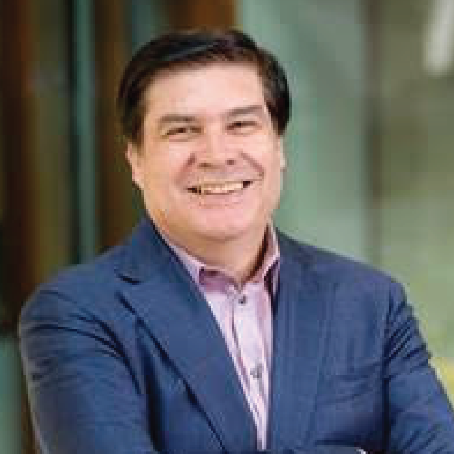 “Micro and Nano Technologies for Cellular Engineering and Material Discovery”
Abstract
The ability to control fluidic, electrical, and mechanical fields using micro and nanotechnologies is enabling key advances in cellular engineering and material discovery. In this presentation, I will address two examples: i) the role of microfluidic platforms in genome engineering and temporal cell analysis, and ii) the use of microsystems for in-situ electron microscopy characterization of low dimensional materials. In cellular engineering, a critical step is the intracellular delivery of gene editing machinery and subsequent nondestructive, temporal analysis of cells. Both can be achieved by cell membrane permeabilization, as part of workflows employed in the investigation of molecular mechanisms of disease, pharmacological screening, and development of new therapeutics. Specifically, I will discuss several microfluidic platforms created in my lab, ranging from a fully automated nanofountain probe electroporation (NFP-E) system, which provides single- cell manipulation with superior cell viability and efficiency, and a live cell analysis device (LCAD) for nondestructive and temporal cellular analysis. A critical aspect of the technology is the possibility of perturbing cell state and downstream pathways. I will present single cell RNA sequencing data analysis to show that microfluidic technology leads to significantly reduced cell stress response and much higher control of molecular payload when compared to standard delivery methods. In a second example, I will discuss microsystems to investigate size scale effects on the mechanics of 1D and 2D materials. These materials are being employed in the development of next- generation electronics, optical, and sensor technologies, as well as in energy production and storage techniques, e.g., supercapacitors, solar cells, and battery electrodes. Such applications involve frequent mechanical deformations such as stretching and bending, so the lifespan (integrity and reliability) of the material is a critical feature. In this context, I will present in situ electron microscopy experiments and atomistic models we have used to understand deformation and failure modes in metallic nanowires and transition metal dichalcogenides. Implications on the development of atomistic models with predictive capabilities, in the spirit of the materials genome initiative, will be highlighted.
Biography
Horacio D. Espinosa is the James and Nancy Farley Professor of Manufacturing and Entrepreneurship, Professor of Mechanical Engineering, and the Director of the Theoretical and Applied Mechanics Program at the McCormick School of Engineering, Northwestern University. He received his Ph.D. in Solid Mechanics from Brown University in 1992. Espinosa has made contributions in the areas of deformation and failure of materials, design of micro- and nano- systems, in situ microscopy characterization of nanomaterials, and microfluidics for single cell manipulation and analysis. He has published over 300 technical papers on these topics. Espinosa received several awards including the Prager Medal from the Society of Engineering Science, the Society for Experimental Mechanics Murray and Sia Nemat Nasser Medals, and the ASME Drucker and Thurston awards. He is a member of the National Academy of Engineering (NAE), foreign member of Academia Europaea, the European Academy of Arts and Sciences, the Russian Academy of Engineering, and Fellow of AAAS, ASME, SEM, and AAM. He was the President of the Society of Engineering Science in 2012 and is a member of the IUTAM General Assembly.
Edward Botchwey, Ph.D.
Professor
The Wallace H. Coulter Department of Biomedical Engineering
Georgia Institute of Technology
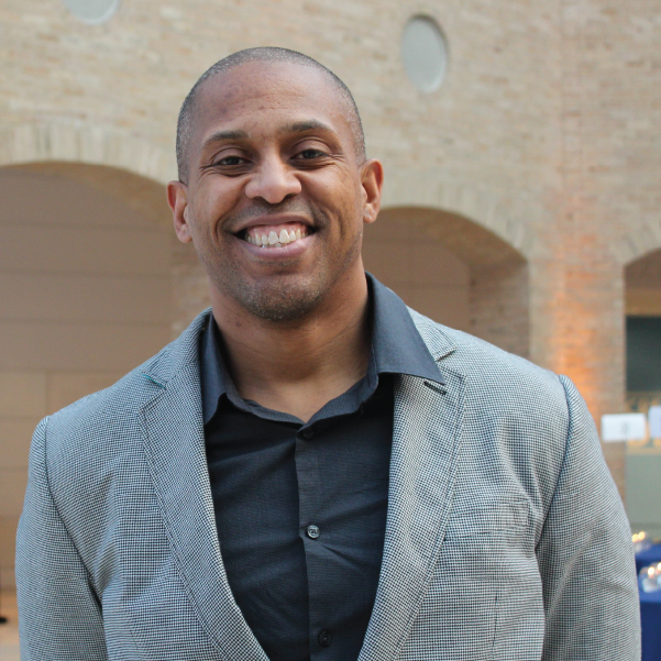 “Unlocking the Power of Bioactive Lipid Signaling for Immunoengineering and Regenerative Medicine”
Abstract
Innate immune cells play a crucial role in tissue growth, stability, and injury recovery. They guide blood vessel restructuring, activate local stem and progenitor cells, and direct tissue repair, such as in muscle and skin. To leverage this innate ability, immunoregenerative biomaterials are being developed that target specific subpopulations of monocytes/macrophages to promote repair by controlling their recruitment, positioning, differentiation, and function in damaged tissues. Monocyte and macrophage phenotypes range from inflammatory (M1) to pro-regenerative (M2) and their functions are heavily influenced by the injury environment. It's believed that dividing labor among different subpopulations of monocytes and macrophages could optimize regenerative functions over inflammatory ones. However, the delicate balance between necessary inflammatory and regenerative functions of myeloid cells is not yet fully understood. This talk will examine distinct populations of monocytes and macrophages, their role in various injury environments, and smart biomaterial design strategies that harness the innate inflammatory response to improve healing.
Biography
Edward Botchwey received a B.S. degree in Mathematics from the University of Maryland at College Park in 1993 and both M.E. and Ph.D. degrees in Materials Science Engineering and Bioengineering from the University of Pennsylvania in 1998 and 2002, respectively. He is currently a Professor in the Wallace H. Coulter Department of Biomedical Engineering at Georgia Tech and Emory University. Dr. Botchwey is a former Ph.D. fellow of the National GEM Consortium, a former postdoctoral fellow of the UNCF-Merk Science Initiative, and a recipient of the Presidential Early Career Awards for Scientists and Engineers from the National Institutes of Health and the Society for Biomaterials Mid-Career Award. Dr. Botchwey’s research focuses on how transient control of immune response using bioactive lipids can be exploited to control the trafficking of stem cells, enhance tissue vascularization, and resolve inflammation.
Juliane Nguyen, Ph.D.
Division of Pharmacoengineering and Molecular Pharmaceutics
Eshelman School of Pharmacy
University of North Carolina at Chapel Hill
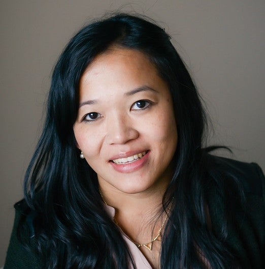 “Engineering Non-Invasive Drug Depots and Auxetic Patches for Cardiac Applications and Dynamic Organ Repair”
Abstract
Dr. Nguyen’s talk introduces an advanced approach to creating non-invasive therapeutic depots that use zipper-mediated crosslinking. She discusses its use in engineering frameworks for creating scaffold-free delivery methods for cardiac repair. Unlike conventional drug depots, this new generation of drug depots can be administered and removed non-invasively. Thus, they do not require surgery. Furthermore, Dr. Nguyen will discuss the development of auxetic patches for dynamic organ repair. Current patches do not conform to the complex mechanics of dynamic organs, such as the lung or heart. To address these limitations, Dr. Nguyen’s lab has developed a scalable patch platform that is instantly adhesive and able to conform to volumetric changes for dynamic organ and wound repair.
Biography
Juliane Nguyen, Ph.D., is an Associate Professor and Vice Chair in the Division of Pharmacoengineering and Molecular Pharmaceutics, School of Pharmacy, at the University of North Carolina at Chapel Hill. Her lab develops complex biologics for treating myocardial infarction, cancer, colitis, and other diseases. Dr. Nguyen’s work has been recognized with the NYSTAR faculty award, the NSF CAREER Award (2018), the Young Innovator Award from the Biomedical Engineering Society (2019), and the AAPS Emerging Leader Award (2019). She received her Ph.D. in Pharmaceutical Sciences from the Philipps-University of Marburg (Germany), where she was mentored by Dr. Thomas Kissel. She then trained at UCSF under Dr. Frank Szoka, where she was a Deutsche Forschungsgemeinschaft Postdoctoral Fellow. Dr. Nguyen is currently a standing member of the NIH Drug and Biologic Therapeutic Delivery (DBTD) study section. She is also the Executive Editor of Advanced Drug Delivery Reviews.
Arvind P. Pathak, Ph.D.
Professor
Depts. of Radiology, Biomedical and Electrical Engineering
The Sidney Kimmel Comprehensive Cancer Center
Institute for NanoBioTechnology
Institute for Computational Medicine
The Johns Hopkins University School of Medicine
 “IMAGE-BASED” SYSTEMS BIOLOGY - The Next Frontier in Bioengineering”
Biography
Dr. Pathak (www.pathaklab.org) is a biomedical engineer, ideator and mentor whose mission is to “improve lives via the transformative power of images”. He received a BS in Electronics Engineering from the University of Poona, India. He received his PhD from the joint program in Functional Imaging between the Medical College of Wisconsin and Marquette University, where he was a Whitaker Foundation Fellow. He completed a postdoctoral fellowship in Molecular Imaging in the Dept. of Radiology at the Johns Hopkins University School of Medicine. At Johns Hopkins University (JHU), Dr. Pathak is Professor in the Schools of Medicine (Radiology and Oncology) and Engineering (Biomedical and Electrical Engineering). He is also a member of the Sidney Kimmel Comprehensive Cancer Center, the Institute for NanoBioTechnology (INBT), and the Institute for Computational Medicine (ICM). As globally recognized expert on functional and molecular imaging, his research has been recognized by multiple awards such as the Bill Negendank Award from the International Society for Magnetic Resonance in Medicine (ISMRM) given to “outstanding young investigators in cancer MRI” and the Career Catalyst Award from the Susan Komen Breast Cancer Foundation. Dr. Pathak has served in multiple leadership roles spearheading imaging initiatives, mentoring junior faculty, review panels for national and international funding agencies, and journal editorial boards. He was the Chair of the Cancer Section for the International Society for Magnetic Resonance in Medicine, serves on the Executive Council of the Microcirculatory Society (MCS) and was the Chair of the NIH’s Emerging Imaging Technologies in Neuroscience (EITN) study section. He directs an interdisciplinary, nurturing and continuously funded laboratory, and has mentored almost a 100 award- winning (e.g. Fulbright and Howard Hughes Fellows) students. He has served on multiple diversity initiatives and is dedicated to mentoring the next generation of thought leaders in bioengineering. He was the recipient of the ISMRM’s Outstanding Teacher Award, was a JHU Career Champion nominee, and recognized as a 125 Hopkins Hero by the School of Medicine for his “outstanding dedication to the core values of Johns Hopkins University”.
Sang Woo Seo, Ph.D.
Associate Professor
School of Chemical and Biological Engineering
Seoul National University, South Korea
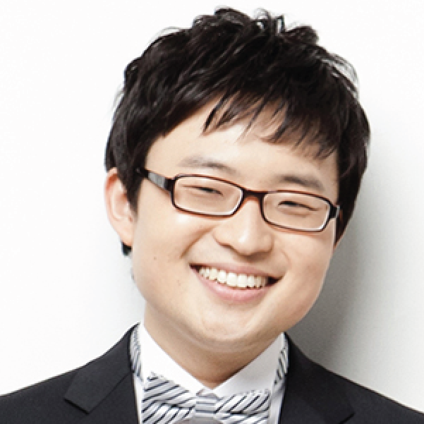 “Microbial Synthetic Biology”
Abstract
The growing interest in the bio-based industry as an alternative production route has given rise to numerous efforts to redesign existing biological systems or synthesize new ones. Imagined to build a system, we require various molecular parts/tools and also need insights on assembling the subset or entire biological systems to exhibit specific functions. The former can be achieved by “Synthetic Biology” approach (bottom-up), and the latter can be achieved by “Systems Biology” approach (top-down). Coordinating these two approaches is mandatory to achieve goals effectively. In this talk, I will describe recent efforts to develop synthetic biology tools such as selection-free tunable plasmid (STAPL) system, synthetic protein quality control (ProQC) system, and biosensor-based adaptive laboratory evolution (ALE) system. Furthermore, their applications in designing and optimizing synthetic cell factories, including newly isolated fast-growing Vibrio sp. dhg to convert marine biomass will be described.
Biography
SangWoo Seo is an Associate Professor in the School of Chemical and Biological Engineering at Seoul National University (SNU), South Korea. He earned a B.S. in Chemical Engineering from POSTECH (Pohang University of Science and Technology) in 2007 and a Ph.D. in Chemical Engineering from POSTECH in 2012, where he worked with Prof. Gyoo Yeol Jung to develop synthetic biology tools in E. coli. He was a postdoctoral scholar in the Department of Bioengineering at UCSD (Prof. Bernhard Palsson’s lab), where he applied multi-omics analyses to reconstruct transcriptional regulatory networks in E. coli. At SNU, his research group focuses on i) elucidation of complex regulatory networks in microorganisms by using next-generation sequencing (NGS) based multi-omics analyses, ii) development of novel synthetic biology tools, and iii) redesigning microorganisms for the production of chemicals, fuels, food additives, pharmaceuticals, and therapeutics, for environmental bioremediation, and for biomedical applications. Several microorganisms such as E. coli (including Nissle 1917), Vibrio natriegens, Corynebacterium glutamicum, Bacillus megaterium, Methanotrophs, Lactobacillus, Saccharomyces cerevisiae, Yarrowia lipolytica, and Pichia pastoris are used as host systems.
Audrey K. Bowden, Ph.D.
Dorothy J. Wingfield Phillips Chancellor Faculty Fellow
Faculty Head, Nicholas S. Zeppos College
Associate Professor, Biomedical Engineering
Associate Professor, Electrical and Computer Engineering
Affiliate Faculty, Vanderbilt Institute of Global Health
Affiliate Faculty, Vanderbilt Institute for Surgery and Engineering
 “New Diagnostic Tools to Enhance the Sensitivity and Specificity of Detection for Early-Stage Bladder Cancer”
Abstract
Bladder cancer (BC) — the 4th most common cancer in men and the most expensive cancer to treat over a patient’s lifetime — is a lifelong burden to BC patients and a significant economic burden to the U.S. healthcare system. The high cost of BC stems largely from its high recurrence rate (>50%); hence, BC management involves frequent surveillance. Unfortunately, the current in-office standard-of-care tool for BC surveillance, white light cystoscopy (WLC), is limited by low sensitivity and specificity for carcinoma in situ (CIS), a high-grade carcinoma with high potential to metastasize. Early detection and complete eradication of CIS are critical to improve treatment outcomes and to minimize recurrence. The most promising macroscopic technique to improve sensitivity to CIS detection, blue light cystoscopy (BLC), is costly, time-intensive, has low availability and a high false-positive rate. Given the limitations of WLC, we aim to change the paradigm around how BC surveillance is performed by validating new tools with high sensitivity and specificity for CIS that are appropriate for in-office use. In this seminar, I discuss our innovative solutions to improve visualization and mapping of suspicious lesions as well as to create miniature tools for optical detection based on optical coherence tomography (OCT). OCT and its functional variant, polarization-sensitive OCT, can detect early-stage BC with better sensitivity and specificity than WLC. We discuss the critical technical innovations necessary to make OCT and PS-OCT a practical tool for in-office use, and new results from recent explorations of human bladder samples that speak to the promise of this approach to change the management of patient care.
Biography
Audrey K Bowden is the Dorothy J. Wingfield Phillips Chancellor Faculty Fellow and Associate Professor of Biomedical Engineering (BME) and of Electrical and Computer Engineering (ECE) at Vanderbilt University. Prior to this, she served as Assistant and later Associate Professor of Electrical Engineering and Bioengineering at Stanford University. Dr. Bowden received her BSE in Electrical Engineering from Princeton University, her PhD in BME from Duke University and completed her postdoctoral training in Chemistry and Chemical Biology at Harvard University. She is a Fellow of SPIE, a Fellow of AIMBE, a Fellow of Optica and a recipient of numerous awards. She currently serves on the Board of Directors of SPIE. Her research interests include biomedical optics (particularly optical coherence tomography and near infrared spectroscopy), microfluidics, and point-of-care diagnostics.
Shreya Raghavan, Ph.D.
Assistant Professor, Biomedical Engineering
Texas A&M University
Adjunct Assistant Professor, Nanomedicine
Houston Methodist Research Institute
 “Metastatic cancer microenvironments: intersecting roles of biomaterials, immunobiology and mechanics”
Abstract
A majority of primary tumors are detectable early, removable via surgery, or treatable with chemotherapy. However, tumor recurrence, metastasis, and chemoresistance are significant causes of mortality in many tumor types. Like any other organ in the body, tumors harbor cancer stem cells, capable of regenerating the tumor or allowing it to escape a chemotherapy insult. Cancer stem cells are also responsible for aggressive metastasis and chemoresistance. Typical to organogenesis and regeneration, tumors actively recruit supporting stromal cells, bypass immune targeting, and recur and metastasize by harnessing microenvironmental cues. Within this context, how cancer stem cells evade immune suppression and metastasize is of significant interest. Unpacking this interaction will lead to the development of new targeted and immunotherapies to curb metastatic spread and its associated mortality. Here, we will look through a biomedical engineer’s lens to identify variables within the cancer stem cell/immune axis, construct engineered models to isolate the variable and further our understanding of how cancers metastasize.
Biography
Dr. Raghavan is an Assistant Professor in the Department of Biomedical Engineering at Texas A&M University. She has a PhD in Biomedical Engineering from Wake Forest University/Virginia Tech, and was a Ruth L. Kirchstein National Research Service Postdoctoral Fellow in Cancer Tissue Engineering at the University of Michigan. At A&M, Dr. Raghavan directs the Stem Cell, Cancer and Immune Tissue Engineering lab, where she builds engineered micro-tissues and micro- environments to study how cancers grow and disseminate. Her approaches integrate mechanobiology, biomaterials and microenvironment engineering to ask questions that intersect the cancer stem cell/immune axis. Her work is funded by the NIH/NCI through an R37 MERIT award, the Department of Defense, and the state cancer agency, Cancer Prevention and Research Institute of Texas. She is also an award winning teacher, recognized for her inclusive pedagogy in the undergraduate classroom by a Montague Scholars Award from Texas A&M University. Dr. Raghavan is an advocate and ally for justice and equity, diversity and inclusion and works actively towards dismantling systemic processes that hold academics behind in STEM. She serves on the DEI Committee of the Society for Biomaterials, to uphold the values of the society to promote equity for all its members.
Viola Vogel, Ph.D.
Professor of Applied Mechanobiology
Department of Health Sciences and Technology
ETH Zürich, Switzerland
 “Mechanoregulation of Cell Niches in Health and Disease”
Abstract
Organ-specific cell niches are not composed of cell monocultures, but of a large variety of cell types which are in close contact to each other and communicate via biochemical and physical factors. Much progress has been made in the molecular understanding of how physical stimuli are sensed and transduced by cells into biochemical signals (mechano-signaling and mechano-transduction), which then regulate gene transcription processes and subsequently cell decision making. Beyond the physical properties of cellular microenvironments, that engineers could easily produce and tune, the reciprocal mechanical signaling between cells and their extracellular environments also involves the stretching of proteins and thus an off-on, or on-off switching of exposed molecular binding sites. Protein stretching can actually switch the structure-function relationships of extracellular as well as of intracellular proteins, which defines the underpinning principles of mechanobiology. Translating what has been learned in mechanobiology (mostly on single cells) to real organs, and finally to the clinic, is hampered by at least two challenges: the lack of nanoscale sensors to probe forces or tissue fiber tensions in healthy versus diseased organs. To bring our knowledge to the patient, we developed a nanosensor to visualize the tensional states of ECM fibers in animal models and in human tissues, with many unexpected findings that were not anticipated from 2D cell culture studies. Underpinning mechano-regulated mechanisms and the significance of these findings in cancer will be discussed.
Francesca Taraballi, Ph.D.
Assistant Professor of Orthopedic Surgery
Director, Center for Musculoskeletal Regeneration
Houston Methodist
 “Immunoengineering solutions for Translational Medicine”
Abstract
The application of immune engineering in translational medicine has emerged as a transformative approach with the potential to revolutionize disease treatment and personalized healthcare. This interdisciplinary field merges principles of immunology, engineering, and nanotechnology to develop innovative strategies for manipulating the immune system to combat a wide range of diseases. Immune engineering has shown remarkable promise in targeted drug delivery, immunomodulation, and regenerative medicine. By harnessing nanoscale materials and biomaterials, immune engineering enables precise interactions with immune cells and immune-related processes, offering unprecedented opportunities for therapeutic interventions. In this seminar, Dr. Taraballi will present her recent advancements in immune engineering research and its impact on translational medicine application in cancer, regeneration, and infection diseases. She will discuss how nanocarriers and immunomodulatory biomaterials are being designed and tailored to effectively deliver therapeutics to specific disease sites while minimizing off-target effects. Moreover, she will explore how immune engineering is facilitating the development of personalized immunotherapies for cancer and autoimmune disorders. Translational studies focusing on preclinical and clinical trials will be also discussed, highlighting the potential of immune engineering to bridge the gap between bench research and bedside applications. As immune engineering continues to advance, it holds immense promise for reshaping the landscape of translational medicine, ultimately improving patient outcomes, and fostering a new era of precision healthcare.
Biography
Dr. Francesca Taraballi is an assistant professor in Orthopedic Surgery at Houston Methodist Hospital and serves as Director for the Center for musculoskeletal Regeneration affiliated to the Orthopedics and Sports Medicine Department. She is also a faculty member of the Center for RNA therapeutics and have adjuctn position at the Department of Nanomedicine and Neal Cancer Center of the Houston Methodist Hospital. Dr. Taraballi own a master in biochemistry and a Ph.D. in Nanotechnology from the University of Milan- Bicocca.
Dr. Francesca Taraballi works in the field of immune engineering. Her expertise lies in the intersection of nanotechnology, biomaterials, and immunology, where she has made significant contributions to the development of innovative approaches for immune modulation and targeted therapeutics. In her research on immune engineering, Dr. Taraballi focuses on designing and engineering nanoscale materials and platforms that can interact with the immune system in precise and controlled ways. These engineered materials are tailored to either enhance the body's natural immune response or to modulate the immune system to target specific diseases.
One of the key areas of her work involves developing nanocarriers for targeted drug delivery. These nanocarriers are designed to encapsulate therapeutic agents, such as drugs or genetic materials, and deliver them specifically to the desired site of action within the body. By leveraging the immune system's ability to recognize and interact with nanoparticles, Dr. Taraballi's research has resulted in the development of more effective and less toxic therapies for a variety of diseases, including cancer, autoimmune disorders, and infectious diseases. Additionally, she has been actively involved in research related to immunomodulatory biomaterials. These biomaterials are engineered to interact with immune cells and promote specific immune responses, such as promoting tissue repair and regeneration or suppressing excessive inflammation. Such advancements have the potential to revolutionize tissue engineering, regenerative medicine, and immunotherapies. Beyond her scientific contributions, Dr. Taraballi has been an advocate for interdisciplinary collaboration. Her work often involves collaborating with immunologists, clinicians, and materials scientists, fostering a holistic approach to tackle complex biomedical challenges. Overall, Francesca Taraballi's work in immune engineering has significantly advanced the field and holds immense promise for the development of more targeted and personalized approaches to healthcare. Her dedication to harnessing the power of the immune system through nanotechnology has the potential to improve the lives of countless individuals affected by various diseases and conditions.
Gunjan Agarwal, Ph.D.
Professor
Department of Mechanical and Aerospace Engineering
Ohio State University
 “A DDR1-Collagen Story in Vascular Biology”
Abstract
Collagen type 1 is the most abundant extracellular matrix protein in adult tissues. The interaction of cells with collagen occurs via specific receptors and regulates both cell and matrix function. We describe here a two-way cross-talk between collagen and the collagen receptor discoidin domain receptor (DDR1). DDR1 is a receptor tyrosine kinases expressed in a variety of mammalian cells. On one hand DDR1 regulates the collagen fibril structure while on the other hand the fibrillar state of collagen impacts receptor function. An altered collagen fibril structure in turn can affect cell-matrix interactions, which have multiple manifestations in health and disease. We elucidate such an effect in vascular biology, wherein the DDR1 knockout mice exhibited enhanced platelet-collagen adhesion and increased thrombogeneity of the vessel wall. Our investigations also reveal that structural alterations in collagen fibril (accompanied by DDR1 activation) exist in vascular diseases such as aortic aneurysms, which can impact platelet adhesion, vascular calcification and mechanics.
Biography
Dr. Gunjan Agarwal is a Professor in the Department of Mechanical and Aerospace Engineering at the Ohio State University. She received her PhD in Biophysics from the Tata Institute of Fundamental Research in Mumbai, India and came to the US for her post-doctoral training at the Albert Einstein College of Medicine (Bronx, NY) and at Procter and Gamble Pharmaceuticals (Cincinnati OH). After a brief period as a scientist at the Wright Patterson Air Force Base, she joined the Ohio State University in 2003.
Prof. Agarwal’s research interests lie “outside the cell” on extracellular matrix remodeling, with a particular focus on the collagen receptors discoidin domain receptors (DDRs). Like integrins DDRs are ubiquitously expressed and are understood to be important for several diseases such as cancers and fibrosis. Dr. Agarwal has been a pioneer in the field of DDRs and her laboratory has yielded seminal work on collagen fibrillogenesis and cell-matrix interactions. Her ongoing research is directed to unravel the causes and consequences of an altered collagen fibril structure with a particular emphasis in vascular and bone diseases.
Prof Agarwal extensively employs atomic force microscopy (AFM) and other microscopy approaches for her research. She directs a multi user AFM core facility and is the co-director of the interdisciplinary Biophysics graduate program at the Ohio State University. She has published 4 book chapters and over 55 journal articles in well-reputed journals like Acta Biomaterialia, J. Mol. Biol, Small etc. Her research has been continuously funded by the NSF, NIH and the American Heart Association. Dr. Agarwal has also been recognized for her efforts in diversity and inclusion.
Kimani C. Toussaint, Jr., Ph.D.
Thomas J. Watson, Sr. Professor of Science
Senior Associate Dean for Research and Strategic Initiatives, School of Engineering
Director, Brown-Lifespan Center for Digital Health
Brown University
 “From SHG Microscopy to Space‐Time Optical Fields: Highlights from the PROBE Lab at Brown University”
Abstract
The remarkable versatility of optical microscopy has helped to both elucidate our fundamental understanding of biological systems and enable the manipulation of matter at the nanoscale. This talk will highlight the major projects pursued by the laboratory for Photonics Research of Bio/nano Environments (PROBE). Specifically, we will review some of the quantitative secondharmonic generation (SHG) microscopy techniques pursued by the PROBE lab for assessment of a variety of collagenous tissues, including recent work where we consider collagen fibers as fluidlike. We will also discuss an area of recent interest developing and utilizing space‐time optical fields for diffraction‐free propagation and speckle‐resistance in the presence of turbid specimens. Finally, we will briefly discuss our interests in developing optical‐based physiological sensors that work equitably across skin tones.
Biography
K. C. Toussaint, Jr., Ph.D. is the Thomas J. Watson, Sr. Professor of Science, and a Professor and Senior Associate Dean for Research and Strategic Initiatives in the School of Engineering at Brown University. Prior to joining Brown in 2019, Dr. Toussaint was faculty at the University of Illinois at Urbana‐Champaign for 12 years. Dr. Toussaint directs the laboratory for Photonics Research of Bio/nano Environments (PROBE Lab), an interdisciplinary research group working in the areas of quantitative nonlinear optical imaging techniques, structured light, nano‐optics, and optical health‐monitoring techniques that mitigate bias. He is a recipient of a 2010 NSF CAREER Award, the 2014‐2015 Dr. Martin Luther King, Jr. Visiting Associate Professor at MIT, the 2015 Illinois Dean’s Award for Excellence in Research, the 2017 Illinois Everitt Award for Teaching Excellence, and the 2019 Distinguished Promotion Award. Dr. Toussaint is also a Fellow of Optica, SPIE, and AIMBE, as well as a Senior Member in the IEEE. In addition, he is a member of the Board of Reviewing Editors for Science, and has served as a Guest Editor for PNAS. Dr. Toussaint’s work on equitable health technologies has appeared in popular press, including NPR, Politico, CNN, STAT+, and the Boston Globe.
Hernan Garcia, Ph.D.
Associate Professor of Genetics, Genomics and Development
Department of Molecular & Cell Biology and Department of Physics
University of California at Berkeley
 “From SHG Microscopy to Space‐Time Optical Fields: Highlights from the PROBE Lab at Brown University”
Abstract
Over the last few decades we have largely identified the repressors and activators that shape gene expression patterns in developing embryos and that, in turn, dictate cellular fates. Yet, despite amassing this great reservoir of knowledge, we are still incapable of predicting how the number, placement and affinity of binding sites for these transcription factors in regulatory DNA dictate gene expression patterns in space and time. Achieving such predictive understanding calls for going beyond molecular parts lists and for obtaining the in vivo biochemical information necessary for fueling theoretical models of transcriptional regulation in developing animals.
In this talk, I will show how we are using physics as a “microscope” to uncover the molecular mechanisms by which activators and repressors dictate transcription in space and time in developing animals. Specifically, using novel quantitative tools that we have developed for precision measurements, I will show that most developmental genes are transcribed in stochastic bursts, and that many transcription factors regulate gene expression by modulating the frequency, duration, and/or amplitude of these bursts. We will then engage in an iterative dialogue between theoretical models and quantitative experiments aimed at revealing the mechanisms underlying this control of transcriptional bursting. Our results challenge the textbook picture of activator and repressor action based on stable protein-protein interactions and call for a description of transcriptional control that acknowledges that the nucleus is not a bag of well-mixed transcription factors. Most importantly, our work sets a path forward for reaching a predictive understanding of cellular decision making and demonstrates how a quantitative dialogue between theory and experiment can shed light on biological mechanisms beyond the reach of even the best super resolution microscopes.
Biography
Hernan G. Garcia is an Associate Professor in the Departments of Molecular & Cell Biology and of Physics at UC Berkeley. As a Physical Biologist, his research aims to uncover the quantitative and predictive principles dictating biological phenomena, with particular emphasis on embryonic development. Hernan is a co-author of the textbook Physical Biology of the Cell and has directed several courses at institutions such as the Kavli Institute for Theoretial Physics at UC Santa Barbara, and the Marine Biological Laboratory in Woods Hole, MA.
Jeanne Stachowiak, Ph.D.
Professor
T. Brockett Hudson Professorship in Chemical Engineering
Departments of Chemical Engineering & Biomedical Engineering
The University of Texas at Austin
 “Intrinsic disorder as an organizing principle for membrane biology ”
Abstract
As the gateway for cellular entry and communication, the surface of the cell holds the answers to critical questions in biology and medicine, while simultaneously providing inspiration for engineered materials and systems. What captivates me about membranes is their ability to precisely and rapidly organize themselves in an environment of staggering complexity. Thousands of distinct protein species reside on cellular membranes, yet functional membrane protein complexes can form within seconds in response to diverse stimuli. Traditionally, structured protein assemblies such as vesicular coats and cytoskeletal filaments have been thought to organize membrane surfaces. In contrast, recent work illustrates that networks composed of proteins with a high degree of intrinsic disorder may provide the necessary flexibility to facilitate efficient assembly of functional protein complexes at membrane surfaces. In particular, our recent work has illustrated that a flexible network of disordered proteins helps to catalyze the assembly of endocytic structures at the plasma membrane. Specifically, we find that an intermediate strength of interaction between these proteins, which leads to liquid-like properties at the macro-scale, maximizes the efficiency of endocytic vesicle assembly. Interestingly, the stability of this catalytic network appears to be controlled by the degree of ubiquitination at endocytic sites, providing a potential mechanism for sensing the presence of transmembrane cargo proteins, many of which are ubiquitinated prior to removal from the plasma membrane. More broadly, the idea of a transition between a stochastic, disordered protein network toward a more ordered structure that is capable of deterministic outcomes provides a template for understanding many processes in membrane biology. At present our group is focused on characterizing these “disorder to order” transitions using a diverse toolset, which includes in vitro biochemistry, quantitative imaging of live engineered cells, and collaborations with theorists who work across a range of length scales. Specific areas of our ongoing work include the role of disordered protein networks in organizing the cytoskeleton, and transmembrane coupling of protein condensates as a novel mechanism of information transfer across biological interfaces.
Sean X Sun, Ph.D.
Professor
Department of Mechanical Engineering
Center for Cell Dynamics (CCD)
Institute of NanoBioTechnology
Johns Hopkins University
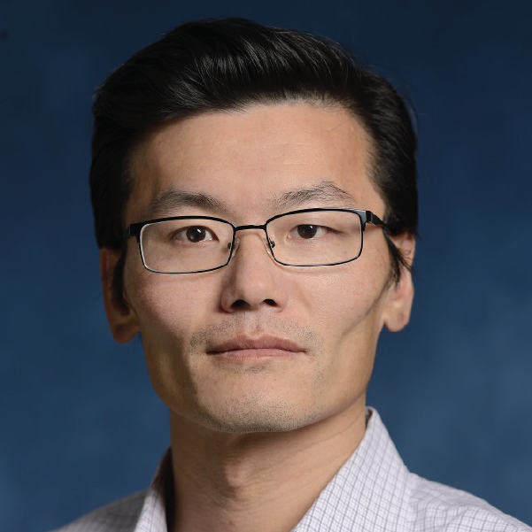 “Water Dynamics in Cells and Tissues”
Abstract
A large fraction of cell and tissue mass is made of water. The flow of water across the cell surface follows osmotic and hydraulic pressure gradients, and is actively controlled by the cell. This physical fact suggests that the mechanical behavior of cells is intimately connected with cell ionic homeostasis and osmotic control. In this talk, we will describe some of our recent experimental and modeling work on water dynamics in cells. We explore how cells control their cytoplasm water content and therefore the cell size. We will describe how cells can use directional flow of water and ion channels such as the Na+/H+ exchanger to propel themselves and migrate, and in the case of epithelia, pump fluid between tissue compartments. In the later case, epithelial pumping of water can strongly influence tissue shape and morphogenesis. Taken together, we suggest a revision of the kinematic law of biological motion that takes these developments into account.
Biography
Sean Sun is a professor of mechanical engineering and biomedical engineering at Johns Hopkins University and a member of the Institute of NanoBioTechnology (INBT) at JHU. His research has been focused on understanding mechanobiology of the cell using experiments as well as mathematical modeling. In the last ten years, he has developed mechanistic mathematical models to understand cell and nuclear size determination in mammalian and bacterial cells, mammalian cell movement and motility, cytoskeleton and water dynamics in moving cells, fundamental processes behind cellular mechanosensation and force generation. He is especially interested in the interplay between mechanics, forces and biochemistry in cells and tissues, and mechanochemical processes in biology. His lab also has developed simple and robust experimental and imaging methods to measure mechanics of biomolecules and live cells. Prof. Sun is a fellow of the American Physical Society and the American Institute for Medical and Biological Engineering.
Camila Hochman-Mendez, PhD
Assistant Investigator
Director, Regenerative Medicine Research and Biorepository Core
The Texas Heart Institute
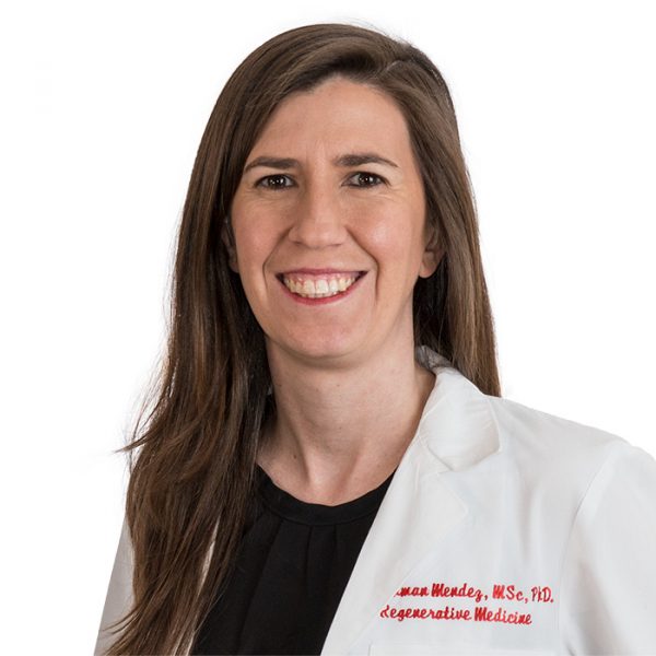 “Innovations in Cardiovascular Tissue Engineering: Addressing Consistency and Reproducibility”
Abstract
The upcoming presentation on cardiovascular tissue engineering will highlight a suite of innovative solutions addressing the critical challenges of consistency and reproducibility in organ regeneration. Through the development of customized bioreactors with coordinated electromechanical stimulation and automated media changes, there's a marked reduction in contamination risks. Incorporating AI-driven technologies for sterile cell injections and the introduction of an in-situ curable semi-conductive hydrogel further propel the field to new heights. Moreover, the strategic application of cardiac dECM powder significantly advances the maturation of hiPSC-derived cardiomyocytes, optimizing their responsiveness to electromechanical stimuli. With the integration of a cutting-edge magnetic bioreactor system and a cost-effective polylaminin protocol, not only is the quality of cardiomyocyte production enhanced, but also its scalability. These advancements represent pivotal strides in the realm of regenerative medicine, emphasizing the potential to revolutionize organ transplantation processes.
Biography
Dr. Camila Hochman-Mendez serves as the Director of the Regenerative Medicine Research Department and is also the esteemed Director of the College of American Pathologists (CAP)-accredited Texas Heart Institute (THI) Biorepository & Biospecimen Profiling Laboratory. With a rich research portfolio, Dr. Hochman-Mendez's work delves into the intricate interplay of extracellular matrix (ECM) proteins in cell-cell interactions, tissue repair, and regeneration. A particular area of fascination lies in understanding the multifaceted interactions between cells and extracellular molecules and vesicles. At THI, the research lab under Dr. Hochman-Mendez's leadership spearheads innovations in bioartificial organ engineering. Notably, the team's efforts in developing customized bioreactors to accommodate and stimulate "rebuilt" organs have garnered significant attention. Their novel approach utilizing decellularized ECM seeded with iPSC-derived cells offers an exciting ECM-based alternative to traditional organ transplantation. A testament to the lab's pioneering work is the publication detailing a fully revascularized rabbit ventricle repopulated with iPSC-derived cardiomyocytes, endothelial cells, and other cardiac cell types. This significant achievement demonstrated the potential of engineered vessels to maintain patency post-transplantation. With the backing of esteemed institutions like the NIH and AHA, Dr. Hochman-Mendez's current research is setting new benchmarks. The team's exploration into micro-controlled multiparametric bioreactors and avant-garde conductive hydrogels for artificial conduction systems is challenging and redefining existing paradigms in regenerative medicine.
Cullen R. Buie, Ph.D.
Associate Professor
Department of Mechanical Engineering
Massachusetts Institute of Technology
 “HIGH THROUGHPUT GENE DELIVERY TECHNOLOGIES FOR APPLIED MICROBIOLOGY AND ENGINEERED CELL THERAPIES”
Abstract
In this talk we will discuss work in our laboratory to use electric fields coupled with fluid flow in microfluidic devices to enable high throughput delivery of nucleic acids to cells. Conventional electroporation approaches for bacterial gene delivery have distinct advantages, but they are typically limited to relatively small sample volumes, reducing their utility for applications requiring high throughput such as the generation of mutant libraries. Here, we present a scalable, large-scale bacterial gene delivery approach enabled by a disposable, user-friendly microfluidic electroporation device requiring minimal fabrication. We demonstrate that the proposed device can outperform conventional cuvettes in a range of situations, including across Escherichia coli strains with a range of electroporation efficiencies, and we use its large-volume bacterial electroporation capability to generate a library of transposon mutants in the anaerobic gut commensal Bifidobacterium longum. Results of this work hold exciting promise for accelerating genetic engineering of bacteria with wide ranging applications in industry and healthcare. Further, we will present recent efforts by a company spun out of the Buie Laboratory, Kytopen, which is leveraging the electroporation work to enable scalable non-viral transfection of mammalian cells. Applications of this work include engineered cell therapies such as CAR-T, which are currently plagued by high costs and manufacturing issues. The non-viral transfection approach developed by Kytopen has the potential to simplify and accelerate the development of life saving cellular therapies.
Biography
Cullen Buie is an associate professor in MIT’s Department of Mechanical Engineering and director of the Laboratory for Energy and Microsystems Innovation. His laboratory explores flow physics at the microscale for applications in materials science and applied biosciences. He earned his master’s and PhD in mechanical engineering at Stanford University and served as a postdoctoral fellow for one year at the University of California-Berkeley. In 2017 Buie co-founded Kytopen, a startup that offers a high throughput method of genetic engineering. Kytopen was among the first start-ups to be backed by The Engine and has raised over $40M in funding to date. Buie has been honored with numerous awards including the NSF Career Award in 2012, the DuPont Young Professor Award in 2013, the DARPA Young Faculty Award in 2013, the NSF Presidential Early Career Awards for Scientists and Engineers in 2016, and in 2021 he was elected Fellow of the American Institute for Medical and Biological Engineering.
Shayn Peirce-Cottler, Ph.D.
Professor and Chair of Biomedical Engineering
Harrison Distinguished Teaching Professor
University of Virginia
 “Combining Experiments with Computational Models to Engineer Tissues”
Abstract
The most prevalent, devastating, and complex diseases of our time, such as diabetes, cardiovascular disease, cancer, and infectious diseases, involve the dynamic interactions of cells with one another and with their changing environment. However, the drugs we typically use to treat diseases target a single protein and disregard the fact that cells within tissues are highly heterogeneous and have individualized responses that contribute to the tissue-level outcomes. To bridge the gap between protein and multi-cell/tissue-levels of spatial scale, my lab develops agent-based computational models and uses them in combination with experiments and machine learning approaches to predict how individual cell behaviors give rise to tissue-level adaptations. We have used agent-based modeling to simulate the structural adaptations of large and small blood vessels, cardiac and skeletal muscle regeneration following injury, and lung tissue remodeling during fibrosis. Our studies have suggested new mechanistic hypotheses and provided guidance for the design of novel therapies that account for the dynamic and heterogeneous interactions between different cell types within diseased and regenerating tissues.
Biography
Shayn Peirce-Cottler, Ph.D. is Harrison Distinguished Teaching Professor and Chair of Biomedical Engineering, with secondary appointments in the Department of Ophthalmology and Department of Plastic Surgery at the University of Virginia (UVA). Dr. Peirce-Cottler received Bachelor’s of Science degrees in Biomedical Engineering and Engineering Mechanics from The Johns Hopkins University in 1997. She earned her Ph.D. in the Department of Biomedical Engineering at the University of Virginia in 2002. Dr. Peirce-Cottler develops computational models and combines them with wet lab experiments to study how tissues heal after injury and to develop therapies for inducing tissue regeneration. She teaches courses in cell and molecular physiology and computational systems bioengineering to undergraduate and graduate students. Dr. Peirce-Cottler has published over 125 peer reviewed papers and book chapters, and she is an inventor on three U.S. Patents. She is a fellow in both the American Institute for Medical and Biological Engineering College of Fellows (AIMBE) and the Biomedical Engineering Society (BMES). She is also Past-President of The Microcirculatory Society. Dr. Peirce-Cottler is a UVA School of Medicine Pinn Scholar, and in 2020 she was awarded the UVA School of Medicine’s Robert H. Kader Award for Excellence in Graduate Teaching and Mentoring. Dr. Peirce-Cottler is passionate about mentoring students and faculty, promoting diversity in STEM, and participating in K-12 outreach to increase students’ interest and self-confidence in pursuing STEM careers.
