Angela K Pannier, PhD
Swarts Family Chair in Biological Systems Engineering
Professor of Biological Systems Engineering and Biomedical Engineering
University of Nebraska-Lincoln (UNL)
 “Engineering Extracellular Vesicles for Nonviral Gene Delivery”
Abstract
This talk will explore the various ways the Pannier Lab is engineering extracellular vesicles for use as nucleic acid carriers, including exosomes derived from mesenchymal stem cells (MSCs) as miRNA carriers, and outer membrane vesicles derived from commensal gut bacteria as oral DNA carriers. For the former, recent studies show that many therapeutic effects of MSCs, a cell type investigated in >1000 clinical trials, are actually mediated by exosomes secreted by MSCs, and these therapeutic effects are mechanistically associated with miRNA delivery, making exosomes derived from MSCs excellent candidates for off-the-shelf miRNA therapies. However, the therapeutic potential of natural exosomes is limited by heterogeneity in miRNA content, meaning therapeutic exosomes could be potentiated by engineering increased loading of specific miRNAs. To this end, we have designed a transgenic system that induces cells to load exosomes with a miRNA of choice, making use of priming strategies we have developed to increase gene transfer to stem cells. For the latter, to overcome challenges associated with oral gene delivery, we are developing a novel, biological-based delivery system by loading outer membrane vesicles (OMVs) derived from commensal gut bacteria with plasmid DNA to create DNA-loaded OMV nanocarriers. OMVs are produced by blebbing of the outer membrane of gram-negative bacteria and act as a natural cell-to-cell communication system for bacteria. OMVs can survive gastric transit and are able to cross the mucosal barrier within the intestine. Additionally, OMVs target epithelial cell via natural host-bacteria interactions and elicit either pro- or anti-inflammatory immune responses depending on from which bacteria they originate, presenting an opportunity to leverage their immunomodulatory properties synergistically with the delivered transgene to tailor oral gene delivery platforms to a variety of applications.
Biography
Dr. Angela Pannier is the Swarts Family Chair of Biological Systems Engineering and Professor of Biomedical Engineering at the University of Nebraska-Lincoln (UNL). Dr. Pannier’s NIH/NSF/USDA/industry supported research focuses on engineering biomaterials and systems for gene therapy and tissue engineering applications. In 2017 she worked as a visiting scholar at the Leibniz-Institut für Polymerforschung in Dresden, Germany. She is an active member of the American Institute of Chemical Engineers, Biomedical Engineering Society (BMES), and the American Society of Gene and Cell Therapy. Dr. Pannier serves as an Associate Editor for Science Advances and on the editorial board for Experimental Biology and Medicine and Regenerative Medicine Frontiers. Dr. Pannier was awarded the 2017 NIH Director’s New Innovator Award for her pioneering work in gene delivery. In 2019 she was awarded a Presidential Early Career Award for Scientists and Engineers (PECASE) from the White House Office of Science and Technology Policy and is the first Nebraskan to earn this honor. She was named a Fellow of BMES in 2020 and AIMBE in 2022. She holds a BS and MS in Biological Systems Engineering from UNL and a PhD from Northwestern University.
Jonathan Rivnay, PhD
Department of Biomedical Engineering
Northwestern University
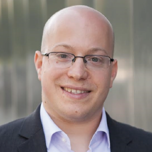 “Polymer-based mixed conductors for applications in bioelectronics ”
Abstract
Direct measurement and stimulation of ionic, biomolecular, cellular, and tissue-scale activity is a staple of bioelectronic diagnosis and/or therapy. Such bi-directional interfacing can be enhanced by a unique set of properties imparted by organic electronic materials. These materials, based on conjugated polymers, can be adapted for use in biological settings and show significant molecular-level interaction with their local environment, readily swell, and provide soft, seamless mechanical matching with tissue. At the same time, their swelling and mixed conduction allows for enhanced ionic-electronic coupling for transduction of biosignals. Through synthetic design and processing we are able to establish design rules towards high performance and stable polymer bioelectronic materials. Such materials open new opportunities for unique form factors and novel device concepts. To this end, I will demonstrate how relaxed design constraints allow for flexible amplification systems for electrophysiological recordings and biochemical detection. Furthermore, advances in additive manufacturing and post functionalization of natural and synthetic biomaterials enable organic bioelectronic composites exhibiting form factors not common of typical bioelectronic devices, which we develop for electroactive scaffolds to modulate tissue state and/or cell fate. These case studies show that fundamental materials research and device engineering continues to fill critical need gaps for challenging problems in bio-electronic interfacing.
Biography
Jonathan Rivnay is an Assistant Professor in the Department of Biomedical Engineering at Northwestern University. Jonathan earned his B.Sc. in 2006 from Cornell University. He then moved to Stanford University where he earned a M.Sc. and Ph.D. in Materials Science and Engineering, studying the structure and electronic transport properties of organic electronic materials. In 2012, he joined the Department of Bioelectronics at the Ecole des Mines de Saint-Etienne in France as a Marie Curie post-doctoral fellow, working on conducting polymer-based devices for bioelectronics. Jonathan spent 2015-2016 as a member of the research staff in the Printed Electronics group at the Palo Alto Research Center (PARC, a Xerox Co.) before joining the faculty at Northwestern in 2017. He is a recipient of the Faculty Early Career Development (CAREER) award from the National Science Foundation (2018), and a research fellowship from the Alfred P. Sloan Foundation (2019). He was recently named a Materials Research Society Outstanding Early Career Investigator (2020), and BMES/CMBE Young Innovator (2021).
Shaoyi Jiang, PhD
Meinig School of Biomedical Engineering
Cornell University
 “Recent Developments in Zwitterionic Materials for Biomedical Applications”
Abstract
An important challenge in many applications is the prevention of unwanted nonspecific biomolecular and macromolecular attachment on surfaces from implants to drug delivery carriers. We have demonstrated that zwitterionic materials and surfaces are highly resistant to nonspecific protein adsorption and microorganism attachment from complex media. Typical zwitterionic materials include poly(carboxybetaine), poly(sulfobetaine), poly(trimethylamine N-oxide), and glutamic acid (E) and lysine (K)-containing poly(peptides). Unlike poly(ethylene glycol) (PEG), there exist diversified zwitterionic molecular structures to accommodate various properties and applications. Furthermore, unlike amphiphilic PEG, zwitterionic materials are super-hydrophilic.
In this talk, I will discuss the application of zwitterionic materials to implants, stem cell cultures, medical devices, and drug delivery carriers. With zwitterionic materials, results show (a) no capsule formation upon subcutaneous implantation in mice up to one year, (b) expansion of hematopoietic stem and progenitor cells (HSPCs) without differentiation, (c) no anti-coagulants needed for artificial lungs in sheep, and (d) no antibodies generated against zwitterionic polymers. I will also discuss recent studies on immunomodulating materials and low-immunogenetic drug delivery carriers and their applications to cancer vaccine and precision medicine.
Biography
Shaoyi Jiang joined the Meinig School of Biomedical Engineering at Cornell as the Robert S. Langer ’70 Family and Friends Professor in June 2020. Before Cornell, he was the Boeing-Roundhill Professor of Engineering in the Department of Chemical Engineering and an adjunct professor of Bioengineering at the University of Washington, Seattle. He received his Ph.D. degree in chemical engineering from Cornell University in 1993. He was a postdoctoral fellow at the University of California, Berkeley between 1993 and 1994 and a research fellow at California Institute of Technology between 1994 and 1996 both in chemistry. He is currently an executive editor for Langmuir (ACS journal), an associate editor for Science Advances (a Science family journal from AAAS), a fellow of the American Institute of Chemical Engineers (AIChE), and a fellow of the American Institute for Medical and Biological Engineering (AIMBE). He received the Braskem Award for Excellence in Materials Engineering and Science, AIChE (2017). His research focuses on biomaterials, drug delivery, cancer vaccine and precision medicine
Emma J Chory, PhD
Post-doctoral Fellow
Collins & Esvelt Laboratories
Massachusetts Institute of Technology
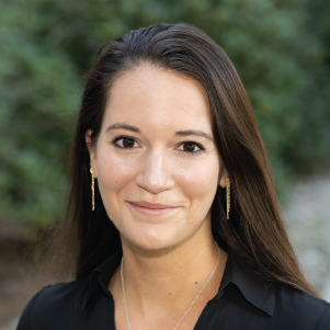 "Phage and Robotics-Assisted Biomolecular Evolution"
Abstract
Evolution occurs when selective pressures from the environment shape inherited variation over time. Within the laboratory, evolution is commonly used to engineer proteins and RNA, but experimental constraints have limited our ability to reproducibly and reliably explore key factors such as population diversity, the timing of environmental changes, and chance. We developed a high-throughput system for the analytical exploration of molecular evolution using phage-based mutagenesis to evolve many distinct classes of biomolecules simultaneously. In this talk, I will describe the development of our open-source python:robot integration platform which enables us to adjust the stringency of selection in response to real-time evolving activity measurements and to dissect the historical, environmental, and random factors governing biomolecular evolution. Finally, I will talk about our many on-going projects which utilize this system to evolve previously intractable biomolecules using novel small-molecule substrates to target the undruggable proteome.
Biography
Emma Chory is a postdoctoral fellow in the Sculpting Evolution Group at MIT, advised by Kevin Esvelt and Jim Collins. Emma's research utilizes directed evolution, robotics, and chemical biology to evolve novel biologic therapies and proteins. Emma obtained her PhD in Chemical Engineering in the laboratory of Gerald Crabtree at Stanford University. She is the recipient of the NSF Graduate Research Fellowship, a pre- and postdoctoral NIH NRSA Fellowship, and recently received the 2022 SLAS Innovation Award for autonomous directed evolution.
Seth Childers, PhD
University of Pittsburgh, Department of Chemistry
 "Protein engineering strategies revealing the functional significance of biomolecular condensates as organizers of the bacterial cytoplasm"
Abstract
One defining difference between bacteria and eukaryotes is the absence of membrane-bound organelles in bacteria. Recently, phase separation of proteins as biomolecular condensates has been recognized as a fundamental way to organize subcellular space as a “membraneless organelle.” Here, we will describe our discoveries of how biomolecular condensates organize and regulate mRNA decay and signal transduction processes in bacteria. Overall, our discoveries further suggest a new understanding of the bacterial cytoplasm as a crowded "bag of biomolecular condensates."
One significant challenge in cell biology is understanding if these membraneless organelles have any functional significance? To consider the function of biomolecular condensates in vivo, the Childers lab engineered a fluorescence biosensor imaging strategy to visualize changes in histidine kinase structure with subcellular resolution. Application of this engineered FRET biosensor visualized that cell pole localized membraneless organelles alter the structure of a critical cell-fate determining protein. Thus, this FRET biosensor strategy provides a strategy to demonstrate the functional significance of biomolecular condensates. It may also be utilized to map signals perceived by two-component systems.
Given our new understanding of the molecular organization of the bacterial cytoplasm, we considered the potential of biomolecular condensate in bacterial synthetic biology. Towards this goal, we revisited the phase properties of a family of ABC triblock peptide nanomaterials that forms hydrogels for drug delivery at high weight percent. We found ABC triblock proteins assemble as gel-like biomolecular condensates at low micromolar concentrations in vitro. Moreover, expression of the coiled-coil triblock peptides in bacteria leads to cell pole accumulation that could sequester clients at the cell pole with the potential to serve as synthetic membraneless organelles in bacteria. In summary, our studies suggest that phase separation allows bacteria to form membraneless organelles that alter our view of bacterial cell biology and present new opportunities in synthetic biology and new antibiotic targets.
Biography
Professor Seth Childers received his B. ChE. degree from the Georgia Institute of Technology in 1999 and worked as a process engineer in the pharmaceutical industry at Merck until 2004. He completed his Ph.D. in biological chemistry at Emory University in 2010 under the mentorship of Professor David Lynn. His graduate studies examined amyloid's phase properties and catalytic capabilities, the causative agent of Alzheimer's Disease. From 2010 to 2015, he was a postdoctoral scholar gaining expertise in bacterial signal transduction in Professor Lucy Shapiro's group at Stanford University. He joined the Department of Chemistry at the University of Pittsburgh in September 2015. His lab has two major research thrusts. One in the area of biomolecular condensates as organizers of biochemistry bacteria. And a second developing area of research is the development of synthetic biology tools to study gut-brain axis dysregulation. Towards this research thrust, the Childers lab applies protein engineering, synthetic biology, microbiology, and biochemistry within each of these research thrusts. He has published 9 peer- reviewed publications, including two papers in Molecular Cell, 2 in ACS Synthetic Biology, 1 in and ACS Sensor. Seth Childers has received two Scialog Fellow Awards as a part of the Chemical Machinery of the Cell 2020-2021 meetings. His research is supported by the NIH and Research Corporation for Scientific Advancement.
Minji Kim, PhD
Postdoctoral Associate
Jackson Laboratory for Genomic Medicine
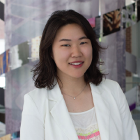 "Molecular communications: transmitting information inside our cells"
Abstract
Our genomes are organized inside the nucleus such that cellular functions are properly performed. This physical structure has been probed by well-established 3D genome mapping technologies Hi-C, ChIA-PET, and their variants, but many questions remain unanswered. I seek to identify who coordinates genome organization, where, when, why, and how. The three protagonists in my story are the zinc finger protein CTCF, a ring-like extruder cohesin, and the transcription machinery RNA Polymerase II. Applying the single-molecule mapping technique ChIA-Drop to human cells revealed that each of these protein factors have distinct roles in forming dynamic loops. In particular, I highlight the dual role of cohesin in forming both structural and regulatory loops, and characterize the causal relationship between genome organization and transcription. These physical communications among protein factors and DNA elements are to be studied further with respect to the time dimension; perhaps the chromatin biology field will expand to study wireless communications in the future.
Biography
Minji is a postdoctoral research associate at the Jackson Laboratory (JAX), studying the relationship between nuclear architecture and gene regulation in human and mouse cells. With BS, MS, and PhD degrees in electrical engineering from UC San Diego (UCSD) and University of Illinois at Urbana-Champaign (UIUC), she develops computational algorithms that enable the analysis of 3D genome mapping data. Her research has been supported by the NIH Pathway to Independence Award and the NSF Graduate Research Fellowship.
Alison Schroer Vander Roest, PhD
Postdoctoral Fellow
Stanford University
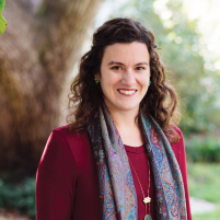 "Cardiac mechanobiology: Modeling altered cardiac biomechanics across scales to understand mechanisms of disease"
Abstract
Heart disease is the leading cause of death in the developed world, and many forms of cardiac disease are characterized by changes in contractile function and tissue stiffness. Hypertrophic cardiomyopathy (HCM) is the most common inherited form of heart disease, and one of the most common sites of disease causing mutations is beta cardiac myosin, the motor protein responsible for contraction. While HCM is characterized clinically by muscle cell (cardiomyocyte) growth and hypercontractility, HCM mutations cause a diverse range of effects on the molecular function of myosin that may have implications for disease severity and treatment options. My postdoctoral research has focused on using gene-edited human induced pluripotent stem cells in engineered environments to measure the effects of these mutations at the cellular scale; namely: increased traction force generation, increased cell size, increased signaling of hypertrophic pathways, and altered cytoskeletal and cell junction organization. I have used computational models to integrate multiple changes in myosin molecular function and cytoskeletal organization to predict changes in cellular forces that can be compared with measured forces to identify driving mechanisms. My research vision leverages engineering tools to clarify the complex mechanical and biological effects of disease-causing mutations and can help inform the development and application of new therapies for HCM and other cardiac diseases.
Biography
Alison Schroer Vander Roest received her undergraduate degree in biomedical engineering at the University of Virginia. She then received a PhD in biomedical engineering at Vanderbilt University with Dr. Dave Merryman, studying fibroblast mechanobiology and the role of cadherin-11 in fibrotic and inflammatory remodeling after myocardial infarction. After completing her doctoral work, Alison pursued postdoctoral training at Stanford University as part of a collaborative team between the mechanical and bioengineering, biochemistry, and pediatric cardiology departments. Her project at Stanford has been co-mentored by Dr. Beth Pruitt, Dr. Jim Spudich, and Dr. Dan Bernstein and aims to understand mechanisms linking mutations in beta-cardiac myosin to phenotypes of hypertrophic cardiomyopathy using stem cell models and engineered environments. Alison has also developed collaborations to incorporate transcriptome analysis, FRET tension sensors, and computational modeling of myosin kinetics to better understand cardiomyocyte mechanobiology. This project has been awarded a K99/R00 Pathway to Independence Award that will fund future research into multiscale mechanisms of cardiac disease.
Collin Tokheim, PhD
Postdoctoral Research Fellow
Harvard T.H. Chan School of Public Health
Dana-Farber Cancer Institute
 "Computationally-driven Identification of Cancer Targets Susceptible to Targeted Protein Degradation"
Abstract
Since their clinical discovery in 2014, Targeted Protein Degradation (TPD) drugs have started to expand the druggable targets in oncology. Unlike existing drugs that block the activity of a target, TPD drugs degrade a protein-of-interest by hijacking the cell’s Ubiquitin-Proteasome System (UPS). However, there is an incomplete understanding about how protein degradation is dysregulated in cancer and how TPD drugs may counteract this effect. Leveraging multi-omics data across more than 9,000 human tumors and 33 cancer types, we found that over 19% of all cancer driver genes impact UPS function. Moreover, we developed a deep learning model (deepDegron) to identify mutations that result in loss of protein degradation signals (degrons), and experimentally validated predictions of point mutations (e.g. in GATA3) and gene fusions (e.g. BCR-ABL) that result in escape from protein degradation. In a second study, we found that altered protein degradation of Cebpd can influence immunotherapy response in a Triple Negative Breast Cancer (TNBC) model by complementing in vivo CRISPR screens with multi-omic data analysis spanning the epigenome, transcriptome and proteome. Lastly, to assess for potential therapeutic vulnerabilities, we developed a machine learning model, MAPD (Model-based Analysis of Protein Degradability), to predict which protein targets are likely degradable by TPD compounds from unbiased proteomic experiments of the kinome. In summary, new computational methods uncovered an underappreciated oncogenic role for dysregulated protein degradation with potential therapeutic implications.
Biography
Collin Tokheim received his Ph.D. in Biomedical Engineering from Johns Hopkins University in 2018. During his Ph.D., his research focused on the development and application of novel computational methodologies to statistically implicate mutations underlying the development or progression of human cancers. He has expertise in machine learning, statistical modeling, genomics, and cancer genetics. Collin is now a Research Fellow at the Harvard School of Public Health and Dana-Farber Cancer Institute. Dr. Tokheim's current research focus is to use computational methods to understand how protein degradation is dysregulated in cancer and how this informs an emerging therapeutic paradigm of Targeted Protein Degradation (TPD) drugs. TPD drugs selectively and potently eliminate -- rather than inhibit -- disease causing proteins. Dr. Tokheim has been the recipient of numerous awards and fellowships, including the Martin & Carol Macht award, Emerging Leaders in Computational Oncology Award, an NCI NRSA fellowship, and the inaugural Damon Runyon fellowship in Quantitative Biology.
Xiao Huang, PhD
Postdoc Scholar
UCSF
 "Precise Modulation of Biological Systems with Advanced Materials"
Abstract
Xiao Huang received his PhD degree in Chemistry and Materials at UC Santa Barbara in 2016. There he developed a method to spatially control biomolecule delivery in human embryonic stem cells mediated by near-infrared light responsive nanocarriers. He is now a postdoc scholar in the lab of Dr. Tejal Desai at UCSF, working at the interface between materials and therapeutic biologics. One highlight from his work is the development of a robust chemistry to surface-functionalize biocompatible materials with exquisite ratiometric precision for applications in immune modulation. Additionally, using advanced imaging methods, he uncovered a new mechanism of epithelial cell tight junction modulation from nanotopographic stimulation. He has published 7 first-authored papers in journals including Nat. Nanotechnol., Adv. Mater., and ACS Nano, etc., is a co-inventor on 2 pending patents, and has been awarded fellowships from UCSF-PBBR, Li Foundation, and CIRM. His future research interest is to develop innovative engineering platforms and analytical methods to advance understanding and modulation of immunological processes.
Biography
Biological systems orchestrate a series of finely controlled events inter- and intracellularly that maintain homeostasis. Synthetic materials can be precisely surface-functionalized with bioactive compounds to modulate cellular events for various biomedical applications. For example, a multi-model display of biomolecules on material surfaces can mimic cell-cell interactions at immune synapses to modulate immunotherapies. Meanwhile, the efficient display of specific functionalities on nanocarriers can facilitate intracellular delivery of biomolecules to regulate or edit genes for disease treatment. Further, the inherent physical features of synthetic materials—size, topography, photoactivity, etc.—provide additional parameters to enable more precise biological control. Currently, there remain several gaps in coordinating the functional capabilities of advanced materials with biological applications. In this seminar, I will describe our work to address challenges in engineering material-cell interfaces. First, we developed a new DNA-scaffold based strategy for biomaterial surface functionalization to allow for density and ratiometric control of multiple functionalities. With this strategy, we designed micron-sized spherical particles with precisely controlled immune modulatory signals and have shown advantages in human T cell activation. We also demonstrated the in vivo utility of this platform through local administration in mice to activate CAR-T cells and minimize off-tumor toxicity. Second, we used surface-functionalized nanoparticles to achieve targeted delivery of biomolecules in cancer and human embryonic stem cells. Importantly, the use of near-infrared light-responsive nanocarriers enabled exquisite cell resolution control of gene silencing guided by light. Third, nanotopographic materials have shown to modulate epithelial tight junctions through a novel mechanism of protein phase separation. Moving forward, I am particularly interested in controlling chemophysical features of functionalized materials to facilitate immunotherapies and gain new knowledge in immunology.
Mahla Poudineh, PhD
Assistant Professor and Director of the IDEATION Lab
University of Waterloo
 "Next-Generation Enabling Technologies for Health Monitoring"
Abstract
To enable personalized and precision medicine, it is crucial to monitor patient health status and bring information on disease-related agents and therapeutic drug molecules into the clinic. This requires new technologies to interrogate different body fluids that are rich sources of biomarkers, such as whole blood and interstitial fluid (ISF). Such technologies enable rapid, sensitive and - ultimately - real-time and continuous analysis of the clinically important biomarkers. Biophysics, materials chemistry and polymer and molecular engineering, as well as micro and nanofabrication, are crucial tools in this endeavor. At IDEATION Lab, we apply innovative engineering solutions to advance patient health monitoring using two main technologies: Microneedles and Microfluidics. In the first part of my talk, I will present our new transdermal biosensing technologies powered by engineered hydrogel microneedles (HMNs), aptamer probes, and in-situ metallic nanoparticle synthesis for minimally invasive, on-needle, and real-time measurement of clinically important biomarkers in ISF. Our HMN assays expect to pave the way for the next-generation of polymeric-based wearable biosensors. In the second part, I will discuss a real-time biosensor driven by microfluidic techniques that continuously updates specific biomolecules’ fluctuating concentration levels with picomolar sensitivity directly in whole blood. For the first time, our microfluidic assay enables measuring the dynamic changes in blood insulin which is an important knowledge gap in diabetes management. The new advances reported in this talk, enrich the level of information that can be collected from different body fluids, and provide new means and potentials for highly accurate patient health status monitoring, thus transforming the field of personalized and precision medicine.
Biography
Mahla Poudineh is an Assistant Professor at the University of Waterloo, Department of Electrical and Computer Engineering and the founding director of IDEATION Lab (Integrated Devices for Early disease Awareness and Translational applicatIONs Laboratory) since January 2020. She received her Ph.D. degree in Electrical Engineering from the University of Toronto in 2016. Prior to joining Waterloo, Mahla completed postdoctoral training at the University of Toronto, and Stanford University, School of Medicine in 2018 and 2019, respectively. Her research interests include developing biosensing approaches for diagnostic and therapeutic purposes, continuous health monitoring and translating biomedical devices to the clinic and market. Her research has been selected as Science Translational Medicine Editor’s choice article and highlighted in the Nature News&Views. She was the recipient of the best poster award during the Micro- and Nanotechnologies for Medicine Workshop held at UCLA (July 2019). She has been also selected as an Inaugural Contributor to the Advanced Materials “Rising Stars” Series and to the Nanoscale Emerging Investigators Themed Collection.
Lei Li, PhD
Postdoctoral Scholar
California Institute of Technology
 "New Generation Photoacoustic Imaging: From benchtop wholebody imagers to wearable sensors"
Abstract
Whole-body imaging has played an indispensable role in preclinical research by providing high-dimensional physiological, pathological, and phenotypic insights with clinical relevance. Yet, pure optical imaging suffers from either shallow penetration or a poor depth-to-resolution ratio, and non-optical techniques for whole-body imaging of small animals lack either spatiotemporal resolution or functional contrast. We have developed a dream machine, demonstrating that a stand-alone single-impulse panoramic photoacoustic computed tomography (SIP-PACT) mitigates these limitations by combining high spatiotemporal resolution, deep penetration, anatomical, dynamical and functional contrasts, and full-view fidelity. SIP-PACT has imaged in vivo whole-body dynamics of small animals in real time, mapped whole-brain functional connectivity, and tracked circulation tumor cells without labeling. It also has been scaled up for human breast cancer diagnosis. SIP-PACT opens a new window for medical researchers to test drugs and monitor longitudinal therapy without the harm from ionizing radiation associated with X-ray CT, PET, or SPECT. Genetically encoded photochromic proteins benefit photoacoustic computed tomography (PACT) in detection sensitivity and specificity, allowing monitoring of tumor growth and metastasis, multiplexed imaging of multiple tumor types at depths, and real-time visualization of protein-protein interactions in deep-seated tumors. Integrating the newly developed microrobotic system with PACT permits deep imaging and precise control of the micromotors in vivo and promises practical biomedical applications, such as drug delivery. In addition, to shape the benchtop PACT systems toward portable and wearable devices with low cost without compromising the imaging performance, we recently have developed photoacoustic topography through an ergodic relay, a high-throughput imaging system with significantly reduced system size, complexity, and cost, enabling wearable applications. As a rapidly evolving imaging technique, PACT promises preclinical applications and clinical translation.
Biography
Lei Li obtained his Ph.D. degree from the Department of Electrical Engineering at California Institute of Technology (Caltech) in 2019. He received his MS degrees at Washington University in St. Louis in 2016. He is currently a postdoctoral scholar in the Department of Medical Engineering at Caltech. His research focuses on developing next-generation medical imaging technology for understanding the brain better, diagnosing early-stage cancer, and wearable monitoring of human vital signs. He was selected as a TED fellow in 2021 and a rising star in Engineering in Health by Columbia University and Johns Hopkins University (2021). He received the Charles and Ellen Wilts Prize from Caltech in 2020 and was selected as one of the Innovators Under 35 by MIT Technology Review in 2019. He is also a two-time winner of the Seno Medical Best Paper Award granted by SPIE (2017 and 2020, San Francisco).
Yvon Woappi, PhD
 "Orchestrating cellular regeneration at organ scale"
Abstract
Large scale tissue damage, such as organ failure and burn injury, is a leading cause of morbidity and death. However, the mechanisms underlying full regeneration of organs remain poorly understood. As the largest organ system in the body, the integumentary system is a composite tissue evolutionarily adapted for healing. Consequently, its complex physiology requires multifaceted cooperation between several distinct cell populations and cell lineages of embryologically distinct origins. Equally integrated within this dynamic process is local immune response that produces mitogenic and inhibitory signals throughout the restoration procedure. There remains a significant gap in understanding how these processes are orchestrated, and how various skin cell populations from distinct developmental lineages functionally cooperate to regenerate tissue at organ scale. My research seeks to characterize the molecular language of tissue healing and to harness this malleable dialect for the regeneration of mammalian organs. Through the development of organoid-based models of wound healing, and the coupling of these systems with novel gene-editing approaches, my work is enabling the functional understanding of the multifaceted cellular events executed throughout restorative healing. Discoveries from this work have established several high throughput technologies and have illustrated their utility in identifying novel regulators of tissue healing.
Biography
Dr. Yvon Woappi’s passion for life sciences ignited during his childhood in Douala, Cameroon and was magnified after his family immigrated to Hanover, Pennsylvania during his middle school years. He went on to receive his B.S in Biology at the University of Pittsburgh, and his Ph.D. in Biomedical Sciences as a Grace Jordan McFadden Fellow under Lucia Pirisi at the University of South Carolina. Dr. Woappi is currently a postdoctoral fellow in the Harvard Dermatology Research Program under the supervision of Matthew Ramsey and Professor Thomas Kupper. His work focuses on engineering 3D skin culture systems and multifunctional gene-editing platforms to study multi-lineage tissue regeneration. He was recipient of the 2019 Engineering The Genome Keystone award and was selected as a Rising Star in Biomedical Sciences by MIT, Cornell, and Columbia University. Most recently Dr. Woappi was awarded the NIGMS-MOSAIC K99/R00 Postdoctoral Career Transition Award to launch his independent research laboratory. Away from the bench, Dr. Woappi is an ardent proponent of inclusive excellence and currently sits on the advisory committee for the NIH Continued Umbrella Research Experiences Program at Harvard Medical School.
Nathan Reticker-Flynn, PhD
Instructor, Department of Pathology
Stanford University
" Roles of systemic immunity in tumor metastasis and effective immunotherapy"
Abstract
The majority of cancer-associated deaths result from distant organ metastases rather than primary tumors, yet few therapies exist to treat this stage of disease. Recent advances in tumor immunotherapies, such as immune checkpoint blockade, have shown promise for patients with metastatic disease, yet most patients remain unresponsive to these treatments. Here, we investigate the roles of systemic immunity in metastatic progression and response to immunotherapies. We demonstrate that lymph node colonization plays a critical role in metastatic progression by imparting tumor-specific immune tolerance within the immune repertoire of the involved lymph nodes. This tolerance becomes systemic across the host and facilitates metastatic seeding of distant sites. Furthermore, using mouse models, mass cytometry, and systems approaches, we demonstrate that the generation of effective anti-tumor immune responses to immunotherapies requires systemic activation of immunity rather than reinvigoration of immune populations within the local tumor microenvrionment. Together, these findings demonstrate the critical roles of systemic immunity in promoting metastatic progression and driving responses to immunotherapy.
Biography
Nathan Reticker-Flynn is a Biomedical Engineer and tumor immunologist working at the interfaces of cancer metastasis, tumor evolution, adaptive immunity, and immuno-oncology. He received his PhD from the Massachusetts Institute of Technology while working with Dr. Sangeeta Bhatia as part of the Harvard-MIT Division of Health Sciences and Technology. His doctoral studies focused upon developing technologies for interrogating interactions between tumors and extracellular matrix during metastatic progression. Using mouse models of lung adenocarcinoma, his studies revealed that metastatic cells alter their adhesion properties, preferentially binding to combinations of ECM containing fibronectin and a family of lectins known as galectins. These interactions are mediated through alterations in integrin expression as well as aberrant glycosylation, the latter of which confers an ability to interact with pro-tumorigenic myeloid populations within the metastatic niche. Dr. Reticker-Flynn performed his postdoctoral studies in the laboratory of Dr. Edgar Engleman at Stanford University School of Medicine. There, his work has focused on using systems approaches and mouse models to investigate tumor-immune interactions during metastasis and responses to immunotherapies. His discoveries include the revelation that effective immunotherapies require systemic activation of anti-tumor immunity and that lymph node metastases serve to reeducate adaptive immune responses in a manner that promotes distant metastasis. His work is funded through various NIH mechanisms as well as a METAvivor Early Career Investigator award.
