After returning to Houston from Costa Rica, the first surgery I observed at the Texas Children’s Hospital was a laparoscopic cystectomy – a procedure to remove an enormous cyst from a 16-year old’s left ovary. Most cysts can be removed using laparoscopy. It is a type of keyhole surgery where small cuts are made in the lower abdomen and gas is blown into the pelvis to allow the surgeon to access the ovaries. Then a laparoscope (a small, tube-shaped microscope with a light on the end) is passed into the abdomen so the surgeon can see the internal organs.
So after collecting my scrubs from a vending machine (yes - now getting your scrub uniform is as easy as getting a bag of chips), I made my way to the OR. The patient had already been sedated and the surgical staff was prepping for surgery. The OR can be a busy place with a lot of unfamiliar technical equipment. As an observer, its best to adopt a fly on the wall approach and stay far away from the sterile field. The anesthesiologist connected the patient to various monitors to keep track of her vital signs and the scrub nurse arranged the sterile instruments to be used during surgery on a stainless steel table. During the surgery, they dimmed the OR lights so that the overhead surgical lights provided brilliant illumination to the surgical site without any shadows. This ambient lighting together with the sound of slow country music made for a very cosy atmosphere in the cold OR.

During laparoscopic procedures, the image is usually displayed on an LCD monitor placed on an instrument tower at eye level, near the operating table. Here, the three-dimensional reality of the body is displayed on a two-dimensional screen. This set-up requires the surgeon to use their hand-eye coordination skills to simultaneously observe the operative field and maneuver the instruments. I stood behind the surgeon and watched the internal organs come to life on this screen.
First, I saw a big, bulging ovary which carried the cyst. The goal was to resect the ovarian wall tissue and remove the cyst. A cystectomy, in many ways, is like peeling an orange. The skin of the orange is the ovarian wall and the edible fruit inside is the cyst. As you slowly peel the skin away from the fruit, it is important to avoid puncturing the water-filled fruit. Similarly, the cyst is also a sac-filled with fluid material and it is vital to not puncture the cyst. Puncturing the cyst could cause the cyst fluid to spill into the uterus and this can complicate the surgery. But this surgery was very successful – they “peeled” the cyst away with minimal damage, the cyst was benign (yay!) and there were no surprises.
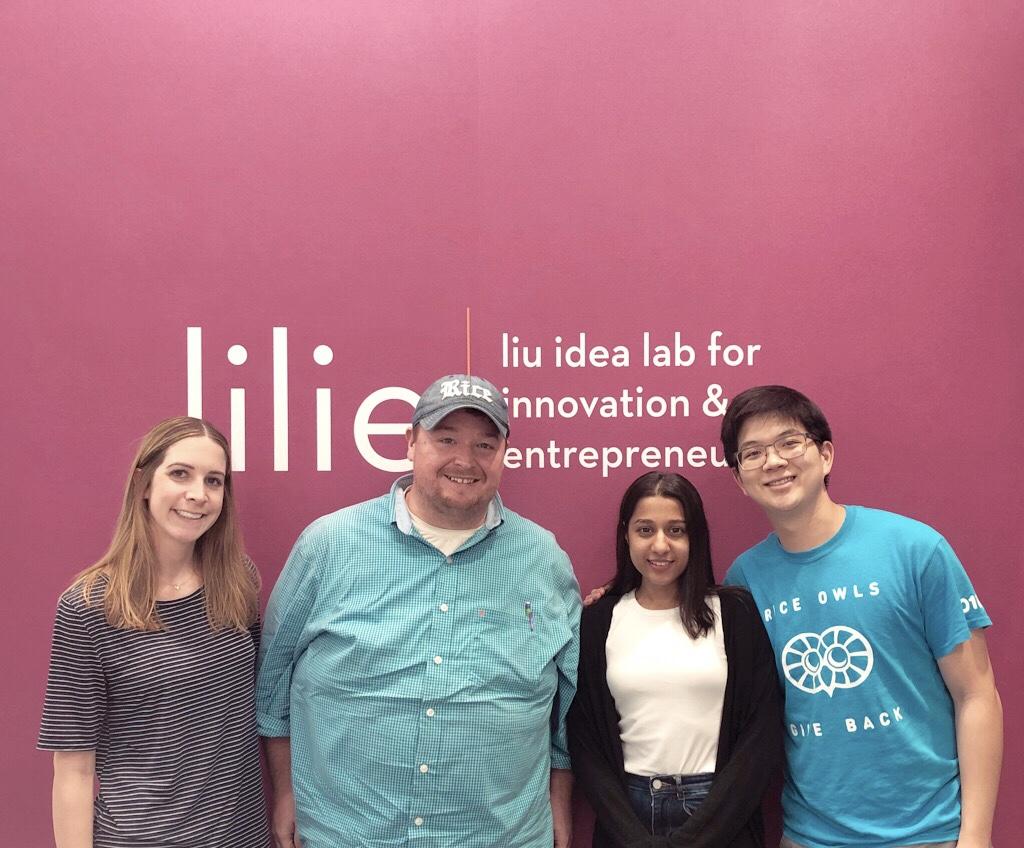
I couldn’t wait to share my observations with my team – Erik, Sydney, Ross and Luke. While Erik and I are in the GMI program, Ross and Sydney are pursuing their masters at the Jones Graduate School of Business and Luke was a practicing physician for ten years. We’ve all been working together to identify unmet clinical needs at the Texas Children’s Hospital in pediatric urology and gynecology and I’m excited to see how our project eventually takes shape in the next few months.
Learn more about our one-year, full-time Master of Bioengineering in Global Medical Innovation.

