
The Rice Bioengineering Colloquia is a seminar series that has a long tradition at the Department of Bioengineering at Rice University. Each week, experts from around the world are brought to Rice to present the latest findings and research in the broadly defined bioengineering field. In Spring 2024, the BIOE Colloquia will be held on Thursdays.
Upcoming Seminars
Thursday, April 25
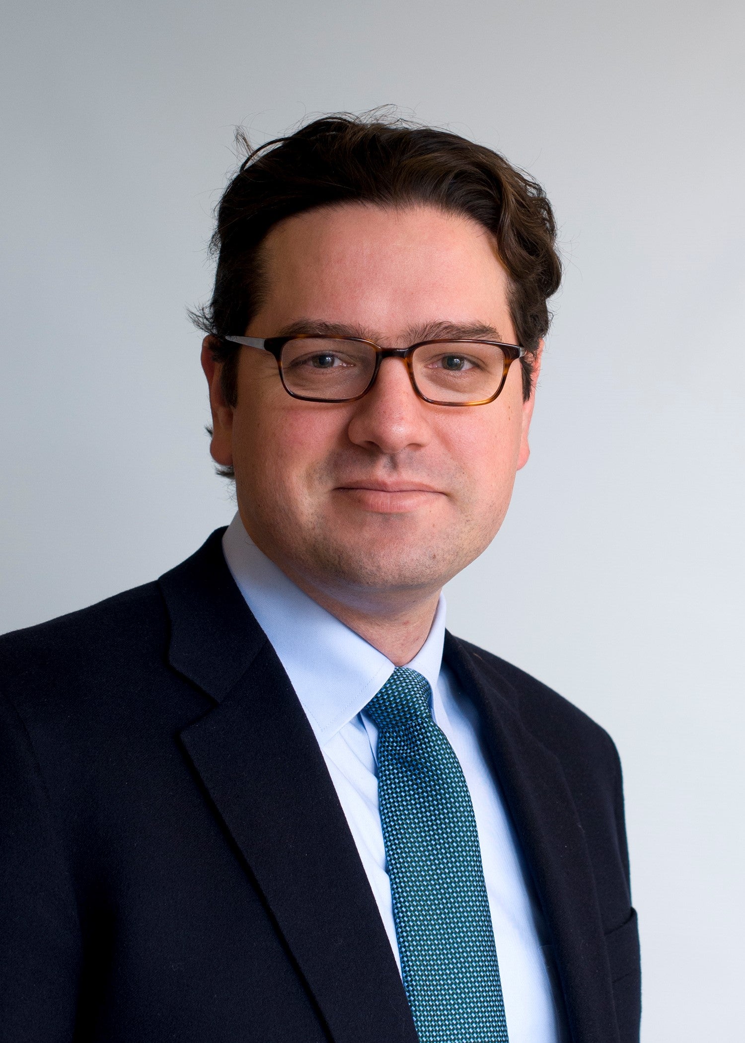 Giovanni Traverso, MB, BChir, PHD
Associate Professor, Department of Mechanical Engineering, Massachusetts Institute of Technology, Division of Gastroenterology, Brigham and Women's Hospital, Harvard Medical School
"Engineering drug delivery and sensing solutions for an extreme environment"
Abstract: Professor Traverso will aim to review ongoing efforts in his lab towards the development of drug delivery and sensing technologies capable of operating in extreme environments like gastrointestinal tract. Specifically, he will present advances in materials science, device development and translational efforts toward addressing medication non-adherence and the dosing macromolecules.
- Biography
-
Dr. Traverso is an Associate Professor in the Department of Mechanical Engineering at the Massachusetts Institute of Technology and Associate Physician in the Division of Gastroenterology, Brigham and Women’s Hospital (BWH), Harvard Medical School. He received his undergraduate and medical degrees from Trinity College, University of Cambridge, UK, and his PhD from the lab of Prof. Bert Vogelstein at Johns Hopkins University where he developed non-invasive tests for the detection of colon cancer. For his post-doctoral research, he worked in the laboratory of Professor Robert Langer at the Massachusetts Institute of Technology (MIT) where he developed a series of novel technologies for drug delivery as well as physiological sensing via the gastrointestinal tract. His current research program is focused on developing the next generation of drug delivery systems to enable efficient delivery of therapeutics through the gastrointestinal tract as well developing novel ingestible electronic devices for sensing a broad array of physiologic and pathophysiologic parameters.
Past Seminars
-
Thursday, April 18
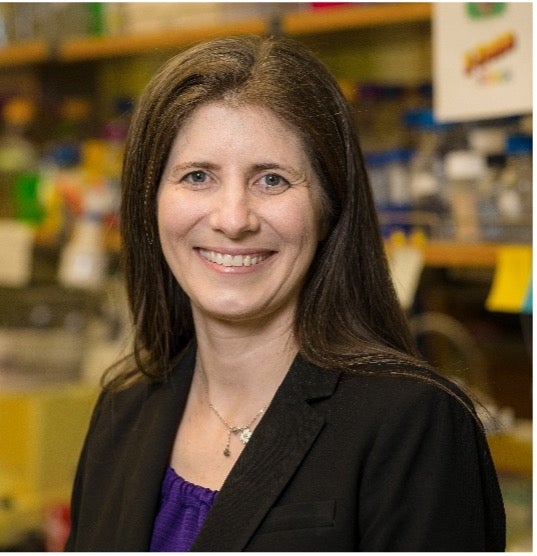
Danielle Tullman-Ercek, PHD
Professor in the Department of Chemical and Biological Engineering at Northwestern University, Co-Director of the NU Center for Synthetic Biology, Director of the Master of Science in Biotechnology Program
“Designing with nanoscale building blocks: How protein engineering enables new solutions for medicine, sustainability, and material”
Abstract:Self-assembling proteins make up precisely ordered nanostructures from filaments and capsules to pumps, each of which, is promising for applications ranging from biomanufacturing to medicine to materials. For example, a nanoscale protein-based container could serve as a vaccine or as a delivery vehicle for cellular or gene therapy. However, such structures must be tunable for each application, and despite great leaps in our ability to predict how amino acid sequences will fold into soluble protein, it remains a significant challenge to predict how proteins come together to form the assemblies and machines that are ubiquitous to life. To address this challenge, and inspired by advances in next-generation DNA synthesis and sequencing, we developed a workflow to fully characterize the assembly competency of all possible single mutations in several model systems, including those from a virus-like particle, a bacterial organelle, and a secretion system. The resulting high-resolution datasets challenge several conventional protein design assumptions on the composition of linkers, mutability of pores, and more. We then used the same approach but screened for desired functions to enhance the performance of each system in its target application space. For example, a protein filament of a secretion system was engineered to confer >2-fold higher production of a target product, a virus-like particle was engineered for improved endosomal release upon sensing a drop in pH, and a bacterial microcompartment was engineered to produce biochemicals in a sustainable manner. With this talk, I will provide examples of how our sequencing-based approach is useful as a tool for uncovering the fundamental rules of self-assembly as well as for engineering new function into self-assembling systems, highlighting how such approaches maybe used to generate nanoscale precision design in next-generation materials
-
- Biography
-
Danielle Tullman-Ercek is a Professor in the Department of Chemical and Biological Engineering at Northwestern University, Co-Director of the NU Center for Synthetic Biology, and Director of the Master of Science in Biotechnology Program. She is also D irector of SynBREU, which is the first NSF-funded Synthetic Biology undergraduate research program. Outside of Northwestern, she was also a founding council member of the Engineering Biology Research Consortium, serves as an Editor for mSystems, and is active in the American Chemical Society Biochemical Technology Division. She is also co-founder of a company, Opera Bioscience, which is built on her research innovations related to biomanufacturing. She received her B.S. in Chemical Engineering at Illinois Institute of Technology in Chicago, and her Ph.D. in Chemical Engineering from the University of Texas at Austin. She carried out postdoctoral research at the University of California San Francisco and the Joint Bioenergy Institute, while part of the Lawrence Berkeley National Laboratory. Tullman-Ercek’s research focuses on building biomolecular devices for a wide range of applications, including energy, materials, manufacturing, and medicine. She is particularly interested in engineering multi-protein complexes, such as virus capsids and the machines that transport proteins and small molecules across cellular membranes. She received several awards for this work, including the Searle Leadership Award, an NSF CAREER award, and the Biochemical Engineering Journal Young Investigator award, and she was inducted as a Fellow of the American Institute of Medical and Biological Engineering in 2023.
Thursday, April 11
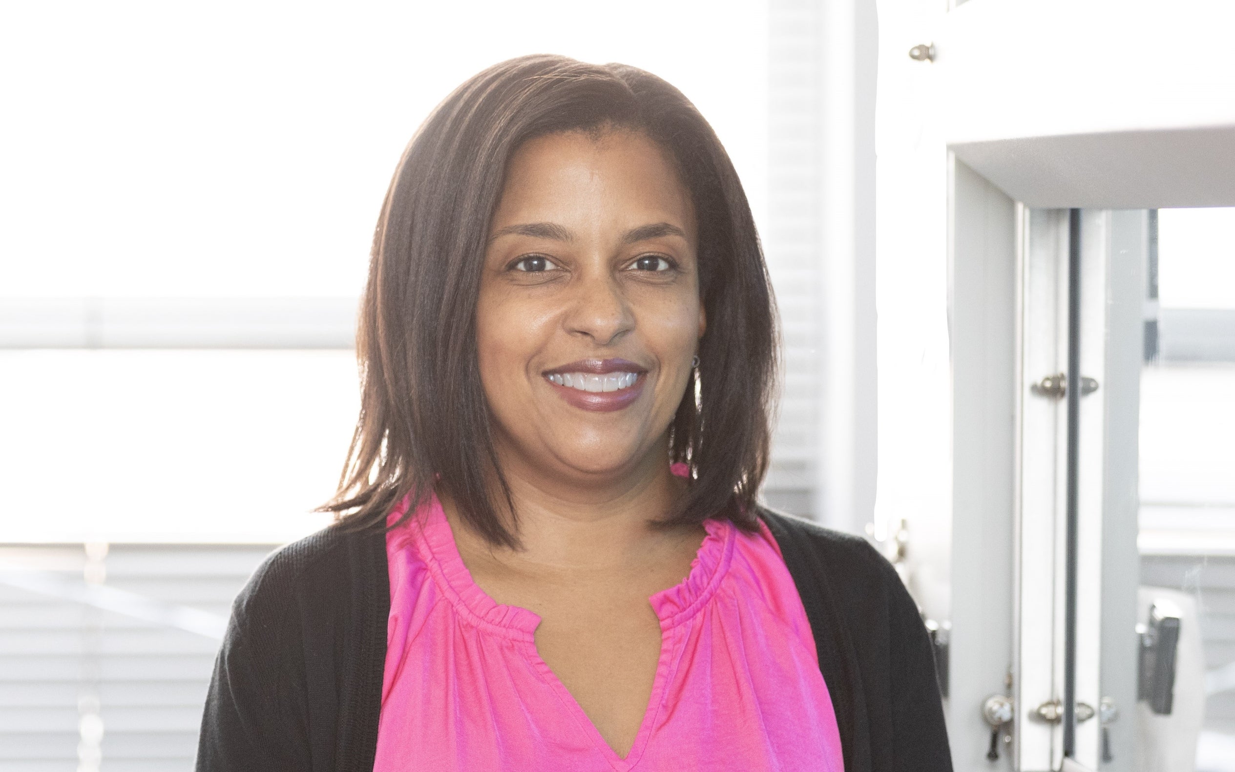 Treena Livingston Arinzeh, PHD
Professor of Bioengineering and Director of the Tissue Engineering and Active Biomaterials (TEAM) Laboratory at Columbia University
"Functional Biomaterials for Tissue Regeneration"
Abstract:
Advances in biomaterial design and its impact on biological function has shown promise in the field of regenerative medicine. This presentation will describe biomaterial properties and designs that impart cues to stem cells and other cell types to affect their behavior and tissue formation in vitro and in vivo. The design of functional, bioinspired materials to be used alone to recruit endogenous cells for tissue repair will also be discussed. Our recent work utilizes protein-based biomaterials as a metabolic approach for bone tissue repair, where studies demonstrate an effect on stem cell migration and differentiation. In combination with a gene knockout model, we are learning about the role of glutamine, which becomes available upon biomaterial degradation, and its effect on bone repair. We have also developed novel glycosaminoglycan (GAG) mimetics, which are sulfated polysaccharides that vary in their degree of sulfation and can be combined to form polymer blends to create scaffolds. Studies demonstrate their sequestration of growth factors and their effect on cartilage repair. ECM proteins, such as collagen and elastin, exhibit electromechanical behavior. Our work using piezoelectric materials, which are materials that provide electrical activity in response to mechanical stimuli, have been explored in in vitro and in vivo models with recent work using degradable, piezoelectric materials having tunable properties. These biomaterials and their potential use in orthopaedic and neural applications will be discussed.
- Biography
-
Treena Livingston Arinzeh, PhD is a Professor of Biomedical Engineering at Columbia University. She is also a co-leader of an Integrated Research Thrust (IRT) and the Director of Diversity of the NSF Science and Technology Center for Engineering Mechanobiology (CEMB). Dr. Arinzeh received her B.S. from Rutgers University in Mechanical Engineering, her M.S.E. in Biomedical Engineering from Johns Hopkins University, and her Ph.D. in Bioengineering from the University of Pennsylvania. She was a project manager at the stem cell technology company, Osiris Therapeutics, Inc. and joined the faculty of the New Jersey Institute of Technology (NJIT) as one of the founding faculty members of the department of Biomedical Engineering. She served as interim chairperson and graduate director, and was promoted to Distinguished Professor in 2020. She joined the faculty of Columbia University in 2022. Dr. Arinzeh has been recognized with numerous awards for her research, including the Presidential Early Career Award for Scientists and Engineers (PECASE). She is a fellow of the American Institute for Medical and Biological Engineering (AIMBE), the Biomedical Engineering Society (BMES) and the National Academy of Inventors (NAI). She has served as chairperson of the National Institutes of Health (NIH) Musculoskeletal Tissue Engineering (MTE) Study Section (2016-2018) and is currently Secretary of the Biomedical Engineering Society (BMES) (2022-2024). She has 16 issued patents and is a co-founder of a start-up medical device company.
Thursday, April 4- **CANCELED**
Hanie Yousefi, PHD
Postdoctoral Fellow, Chan Zuckerberg Biohub, Chicago, Illinois
Future Leader in Bioengineering
"Nanoscale Approaches to Precision Diagnostics and Individualized Medicine"
Abstract: Molecular measurement systems are fundamental to modern medicine. However, these tools provide only a snapshot of a patient's condition and the disease. The emerging field of bioelectronics has the potential to revolutionize healthcare by enabling continuous health monitoring. Devices capable of interacting with the human body, reading and processing information can revolutionize our understanding of disease and enable personalized treatment. Current challenges to the comprehensive integration of molecular diagnostics with bioelectronics include limited long-term functionality, stability, and robustness. State-of-the-art molecular diagnostic tools are often resource-intensive and expensive, rendering them inaccessible to many communities worldwide. My research program is dedicated to addressing these technological shortcomings that have hindered the integration of bioanalytical tools into modern medicine. In this seminar, I will discuss our recent multidisciplinary efforts to develop robust diagnostics tool kits including a reagent-free electrochemical assay for biomarker analysis as well as the engineering of an optical method for monitoring food health. I will then introduce the future plans of the Yousefi lab. We aim to advance alternative manufacturing methods for rapid diagnostics and engineer measurement systems that are resilient, durable, and capable of withstanding harsh conditions. By overcoming bio-interface limitations, I aim to not only introduce long-lasting in vivo function to medical devices but create opportunities for high-throughput, high resolution in vitro disease analysis.
- Biography
-
Doctor Hanie Yousefi (She/Her/Hers) is a biotech innovator, mentor, entrepreneur, and an advocate for equitable health. Hanie is currently a Chan Zuckerberg Fellow building tissue integrated bioelectronics for understanding the role of inflammation in disease progress. She holds a BSc and an MSc in Chemical Engineering. Hanie earned her PhD degree in Pharmaceutical Sciences from the University of Toronto under the supervision of Dr. Shana Kelley where she built reagentless electrochemical systems for infectious disease diagnostics. She co-founded a biotech startup, Arma Biosciences, in 2020 to take this technology to the market. Hanie has mentored over 20 high school, undergraduate, and graduate students and is a proponent of teamwork having published with over 50 co-authors. Hanie has a demonstrated record in service to her community and has held multiple leadership roles such as the chair of student services and the president of the graduate student association in her past institutions. She has been well recognized during her studies with awards and honors such as the NSERC-CGSD PhD fellowship, NSERC-PDF, Pfizer Canada graduate student fellowship and University of Toronto’s leadership award. She has been named a rising star by MIT, Stanford U., and UC Berkeley, a future faculty by ACS PMSE and MRS, and a CAS future leader. She is an advocate for equitable personalized health for everyone and is dedicated to building a research program aimed to build affordable and scalable diagnostics technologies.
Thursday, March 28
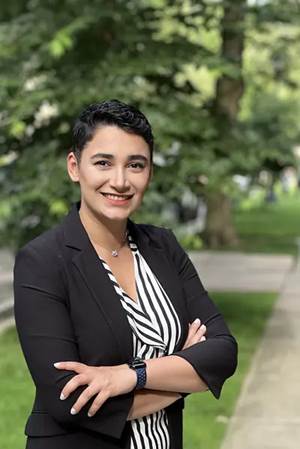
Kharimat Lora Alatise, PHD Candidate
Clemson University
Future Leader in Bioengineering
"Engineering multifunctional peptides to improve nucleic acid delivery for ovarian cancer treatment"
Abstract: RNA interference (RNAi) therapies, such as small interfering RNA (siRNA), are high precision therapies capable of modulating the expression of disease-promoting genes with minimal off-target effects. For aggressive diseases like ovarian cancer, RNAi therapies can serve a robust treatment modality if delivered successfully. Ovarian cancer requires an effective therapeutic strategy to sensitize tumors to chemotherapy due to high relapse rates and drug resistance in advanced stages. As of 2024, the FDA has only approved six siRNA therapies- none of which are approved for cancer treatment. Enhancing siRNA delivery to extrathepatic sites is essential due to the extracellular and intracellular barriers that limit its effectiveness. One promising avenue for overcoming delivery challenges lies in the rational design of amino acid sequences, which can be tailored to engineer peptides with diverse functionalities. These peptides hold the potential to serve as versatile nucleic acid delivery systems, capable of navigating the intricate barriers within the body to reach their intended targets. This talk will highlight the feasibility of peptide sequences to effectively deliver siRNA and silence oncogenes in in vitro and in vivo ovarian cancer models. By harnessing the unique properties of these engineered peptides, potentially new avenues for targeted RNAi therapy can be unlocked, not only for ovarian cancer but also other diseases where RNSi therapy holds therapeutic promise
- Biography
-
K. Lora Alatise earned her B.S. in Biomedical Engineering from the University of Rochester, working with Dr. Danielle Benoit in her Therapeutic Biomaterials lab as a Ronald E. McNair Postbaccalaureate Achievement Scholar. Her research focused on characterizing peptide-conjugated polymeric nanoparticles for small molecule drug delivery. Currently, she is a Ph.D. candidate in Bioengineering at Clemson University, conducting research in Dr. Angela Alexander-Bryant’s Nanobiotechnology Lab, where her dissertation focuses on improving nucleic acid delivery to ovarian tumors. At Clemson, Lora has garnered recognition for her research and service, receiving several awards including the 2023 Eugene M. Langan III Service Award and the Call Me Doctor Fellowship. During her Ph.D., she gained additional expertise by conducting formulation research in Genetic Medicine at Eli Lilly and Company. Upon completing her Ph.D., Lora aims to continue advancing targeted delivery systems for nucleic acid delivery to study and treat diseases that affect distinct and minoritized populations. Lora is passionate about training the next generation of engineers – she actively mentors undergraduate students in research projects and outside of the lab.
Thursday, February 29
Andrew Tsourkas, PHD
Professor and Undergraduate Chair, Department of Bioengineering at The University of Pennsylvania
"Engineering novel antibody and nanoparticle platforms for imaging and therapeutic applications
Abstract: A major goal of my research program is to develop novel molecular imaging agents and targeted therapeutics. In particular, we are (i) developing new nanoformulations for diverse biomedical applications, including image-guided surgery, photodynamic therapy, and sonodynamic therapy; (ii) investigating new targeted strategies that maximize specificity and sensitivity or improve the accumulation and penetration of nanomaterials within tumors; and (iii) developing new bioconjugation techniques that enable the highly efficient, site-specific labeling of proteins, including antibodies. These bioconjugation tools have allowed us to pursue a variety of unique applications, including the conversion of cancer patients' own antibodies into tumor-targeting, T cell re-directing bispecific antibodies. We have also taken advantage of our site-specific bioconjugation technologies to deliver antibodies and proteins into the cytoplasm of living cells, which has enabled the inhibition and degradation of 'undruggable' protein targets. We hope that these tools will help facilitate the movement towards more personalized medicine, whereby treatments are tailored to individual patients.
- Biography
-
Andrew Tsourkas, Ph.D. is a Professor and Undergraduate Chair of Bioengineering at the University of Pennsylvania. He received his Bachelor’s degree in Mechanical Engineering in 1997 from Cornell University and his Ph.D. in Biomedical Engineering from the Georgia Tech/Emory University joint Ph.D. program in 2002. He conducted a post-doctoral fellowship in the Department of Radiology at Harvard University, before joining Penn in 2004. Dr. Tsourkas is currently the Co-Director for the Center for Targeted Therapeutics and Translational Nanomedicine. He was a recipient of the Coulter Foundation Early Career Award, the National Science Foundation CAREER Award, and was elected fellow of the American Institute for Medical and Biological Engineering.
Thursday, February 15
Robert Bowles, Ph.D.
Associate Professor, Department of Biomedical Engineering at the University of Utah
"CRISPR for Discovery, Design, and Development of Musculoskeletal Therapeutics"
Abstract: The development of CRISPR gene regulation systems have opened the door for novel gene and cell therapy strategies. While these systems allow for the targeted and precise regulation of gene expression and can be utilized to engineer cells for a multitude of applications, these systems are also very powerful tools for discovering novel biology and therapeutic targets. Here we will cover its use to neuromodulate pain, engineer cells for tissue engineering in the first part of the talk and explore our discovery of a novel regulator or senescence using theses systems, ZNF865, and its broad potential applications to aging, musculoskeletal disorders, and neurodegenerative disorders.
- Biography
-
I have been studying the intervertebral disc (IVD) and related back pain for over two decades (23 years). I started as an undergrad at the University of Pennsylvania investigating spine biomechanics, which ignited my interest in IVD tissue engineering strategies that target the regeneration and replacement of degenerative IVD tissue at Cornell University. Wanting a better understanding of the underlying mechanisms of pain, I pursued postdoctoral training as an NIH NRSA Postdoctoral Fellow at Duke University investigating the development and maintenance of pain in musculoskeletal preclinical animal models of osteoarthritis and peripheral neuropathies. This postdoctoral training had the added bonus of providing training in lentiviral gene delivery systems, drug delivery for inflammation, and, gene editing/regulation. I have been primarily focused on combining these skill sets, and using CRISPR gene regulation, to solve critical problems in the musculoskeletal space. My laboratory has the distinction of being the first to modify a stem cell with CRISPR epigenome editing and the first to characterize ZNF865, a novel regulator of senescence. My laboratory is currently funded by the NIH to investigate CRISPR epigenome editing in multiple cell types
Thursday, February 22
 Juan Hu, Ph.D.
Postdoctoral Fellow-University of California, Irvine
"High-Throughput Technologies to Illuminate the Molecular Basis of Membrane Penetration within the Rule of 5 and Beyond"
Future Leader in Bioengineering
Abstract: How do molecules cross the cell membrane and what properties allow them to do so? Lipinski’s Rule of 5 generally defines physicochemical properties (molecular weight, lipophilicity) that correlate with good membrane permeability and oral bioavailability. Increasingly, drug discovery projects involve so called “undruggable” protein targets, which generally benefit from chemical space beyond the Lipinski's rule of 5 (bRo5), but these molecules suffer compromised cellular permeability and bioavailability due to unclear structure-permeation relationships. I have developed high-throughput permeation measurement technology – which could elucidate such relationships – as demonstrated through the discovery of unexpected relationships between stereochemistry and membrane permeation. My talk will describe this discovery and its potential implications for drug discovery and the origins of life. I will also discuss our ongoing efforts to explore and define the properties that confer membrane permeability within the Rule of 5 and beyond.
- Biography
-
Dr. Hu achieved her doctoral degree in chemistry from Auburn University in 2018 under the guidance of Professor Christopher J. Easley in the Department of Chemistry and Biochemistry. There, she developed sensitive immunoassays and microfluidic tools for studying dynamics of hormone secretion from cells related to diabetes and obesity. She then joined the Department of Chemistry at Scripps Research for her postdoctoral training under the guidance of Professor Brian M. Paegel, and later moved with the Paegel Laboratory to UC Irvine in 2019. During her postdoctoral training, she has studied membrane permeability and developed high-throughput permeation measurement and screening methods. Her research has been published in Nature Chemistry, Journal of Medicinal Chemistry, Analytical Chemistry, and Lab on a Chip. Dr. Hu received an NIH Pathway to Independence Award (K99/R00).
Thursday, January 25
 Arnab Mukherjee, Ph.D.
Assistant Professor of Chemical Engineering & Biological Engineering at the University of California, Santa Barbara
"Engineering biological water diffusion to create new genetic reporters for molecular MRI"
Abstract: The study of biological functions in intact organisms requires noninvasive genetic reporters to track cells, image gene expression, and monitor signaling pathways. While fluorescent and bioluminescent proteins are widely used as reporters, their utility in deep tissues is limited due to the scattering and absorption of light, which impede imaging beyond a depth of ~ 1 mm from the tissue surface. To overcome this challenge, my research harnesses unexpected connections between proteins and the physics of magnetic resonance (MRI) to create new biomolecular reporters for deep tissue imaging. In this talk, I will discuss our recent efforts to address three long-standing challenges in the development of viable MRI reporters: sensitivity, specificity, and sensor design. First, I will highlight our recent work in increasing reporter gene sensitivity to detect small numbers of genetically labeled cells, potentially, as few as hundred cells per imaging voxel. I will then describe the creation of chemically erasable reporters, which enable “hotspot” imaging with a low tissue background. Finally, I will discuss a new modular approach for programming MRI sensors based on protease modulation of reporter activity.
- Biography
-
Arnab Mukherjee is an Assistant Professor of Chemical Engineering & Biological Engineering at the University of California, Santa Barbara. Prior to arriving at UCSB, Dr. Mukherjee completed a James G. Boswell fellowship in Molecular Engineering at Caltech (working with Prof. Mikhail Shapiro) and obtained his Ph.D. in chemical and biomolecular engineering from the University of Illinois, Urbana-Champaign. The Mukherjee lab works at the intersection of molecular engineering, synthetic biology, and molecular imaging to create new genetic reporters and sensors for magnetic resonance imaging (MRI). Research in the Mukherjee group has been consistently supported by the NIH, Army, and foundations; and recognized with notable awards, including an Outstanding Young Investigator Award (NIH MIRA), a Discovery Award from the DoD, the NARSAD Young Investigator Award from the Brain & Behavior Research Foundation, and a 2022 Scialog Fellows award in Advanced Bioimaging.
Thursday, January 11
 Robert Gray, Ph.D.
Professor of Respiratory Medicine at the School of Infection and immunity and an Honorary Consultant Pulmonologist at NHS GGC
"Cystic Fibrosis lung disease from inflammation to resolution and repair"
Abstract: Cystic Fibrosis (CF) is a genetic disease that is characterized by progressive and destructive lung disease. RG's seminar will cover how inflammation is a key feature of CF and in particular how CF immune cell dysfunction is key feature of the inflammatory process. New CF drug therapies that target the basic defect in CF have additional significant effects on CF immune cells that may be beneficial for people with CF and are increasing our understanding of the inflammatory process in CF. We are now using cells from people with CF to define the on-going inflammation process in CF and deploying them in ex-vivo tissue models to drive therapeutic discovery. A full understanding of the inflammatory process in CF will require cross-disciplinary collaborative research that will allow new therapeutic approaches to be discovered and tested before taking treatments back into clinical trial. RG will outline these initiatives today.
- Biography
-
Robert Gray is Professor of Respiratory Medicine at the School of Infection and immunity and an Honorary Consultant Pulmonologist at NHS GGC. He completed training in Pulmonology in Edinburgh and then held consecutive intermediate (Welcome) and senior (NRS/CSO) fellowships prior to his appointment in Glasgow. The Gray lab studies lung inflammation, damage, and repair processes with a focus on Cystic Fibrosis and related diseases of the airways.
Thursday, January 18-CANCELLED
 Joseph DeSimone, Ph.D.
Sanjiv Sam Gambler Professor of Translational Medicine and Chemical Engineering. Departments of Radiology and Chemical Engineering. Department of Chemistry (by Courtesy). Department of Materials Science & Engineering (by Courtesy). Graduate School of Business (by Courtesy). Stanford University
"The Delicate Interplay Between Light, Interfaces and Design: The Complex Dance that Allows 3D Printing to Scale to Manufacturing"
Abstract: The production of polymer products relies largely on age-old molding techniques. A major reason for this is that additive methods have not delivered meaningful alternatives to traditional processes-until not. In this talk, I will describe Continuous Liquid Interface Productions (CLIP) technology, which embodies a convergence of advances in software, hardware, and materials to bring the digital revolution to polymer additive manufacturing. CLIP uses software-controlled chemistry to produce commercial quality parts rapidly and at scale by capitalizing on the principle of oxygen-inhibited photopolymerization to generate a conjugal liquid interface of uncured resin between a forming part and a printer's exposure window. Instead pf printing layer-by-layer, this allows layerless parts to 'grow' from a pool of resin, formed by light. Compatible with a wide range of polymers, CLIP opes major opportunities for innovative products across diverse industries. Previously unmakeable products are already manufactured at scale with CLIP, including the large-scale production of running shoes by Adidas (Futurecraft 4D); mass customized football helmets by Riddell; the world's first FDA-approved 3D printed dentures; and numerous parts in automotive, consumer electronics, and medicine. At Stanford, we are pursuing new advances including digital therapeutic devices in pediatric medicine, new multi-materials printing approaches, recyclable materials, and the design of a high-resolution printer to advance technologies in the microelectronics and drug/vaccine delivery areas, including novel micro needle designs as a potent vaccine delivery platform.
- Biography
-
Joseph M. DeSimone is the Sanjiv Sam Gambhir Professor of Translational Medicine and Chemical Engineering at Stanford University. He is also Co- Director of Stanford’s Precision Health and Integrated Diagnostics (PHIND) Center (Canary Center) and the founding Faculty Director of the Center for STEMM Mentorship at Stanford. He holds appointments in the Departments of Radiology and Chemical Engineering with courtesy appointments in the Department of Chemistry, the Department of Materials Science and Engineering, and Stanford’s Graduate School of Business. Previously,
DeSimone was a professor of chemistry at the University of North Carolina at Chapel Hill and of chemical engineering at North Carolina State University. He is also Co-founder, Board Member, and former CEO (2014 - 2019) of the additive manufacturing company, Carbon.
DeSimone has published over 380 scientific articles and is a named inventor on over 240 issued patents. He has mentored 80 students through Ph.D. completion in his career, half of whom are women and members of underrepresented groups in STEM. In 2016 DeSimone was recognized by President Barack Obama with the National Medal of Technology and Innovation, the highest honor in the U.S. for achievement and leadership in advancing technological progress.
DeSimone is responsible for numerous breakthroughs in his career in areas including green chemistry, medical devices, nanomedicine, and 3D printing, also co-founding several companies based on his research. In the 1990s he and students invented a green manufacturing process for the synthesis of fluoropolymer materials that eliminated so-called “forever chemicals” like PFAS, which was only partially commercialized by DuPont. In the mid-2000s, DeSimone and students developed a nanoparticle manufacturing platform rooted in an imprint lithography-based r2r process, PRINT (particle replication in non-wetting templates)—the first technology to enable large-scale fabrication of uniform nanoparticles for medicine with independent control over particle features such as size, shape, and composition. Based on PRINT, DeSimone co-founded Liquidia Technologies (NASDAQ: LQDA), which has multiple clinical products. DeSimone’s lab published a large body of research using PRINT to study how specific particle features influence biological processes and to advance the design of vaccines.
More recently, DeSimone and team invented a revolutionary 3D printing technology, CLIP (continuous liquid interface production). CLIP eliminates the slow, layer-by-layer construction seen with other polymer 3D printing approaches to enable parts to ‘grow’ continuously and rapidly from a pool of liquid resin. CLIP delivers production-grade parts comparable in performance to injection molded parts. Based on CLIP, DeSimone co- founded, and was the CEO of for six years, Carbon, Inc., now a global digital additive manufacturing company helping to advance product innovation in numerous industries, including medical, dental, footwear, automotive, and aerospace. CLIP is also used by many academic laboratories to advance research in areas including medical devices and implants.
DeSimone has received numerous recognitions for achievements in science, engineering, invention, and business. In addition to the U.S. National Medical of Technology and Innovation, these include the U.S. Presidential Green Chemistry Challenge Award (1997); the American Chemical Society Award for Creative Invention (2005); the Lemelson-MIT Prize (2008); the NIH Director’s Pioneer Award (2009); the AAAS Mentor Award (2010); the Kabiller Prize in Nanoscience and Nanomedicine (2015); the Heinz Award for Technology, the Economy and Employment (2017); the Wilhelm Exner Medal (2019); the EY Entrepreneur of the Year Award (2019 U.S. Overall Winner); and the Harvey Prize in Science and Techonlogy (2020). He is an elected member of the American Academy of Arts and Sciences one of only 25 individuals elected to all three branches of the U.S. National Academies (Sciences, Medicine, Engineering). DeSimone received his B.S. in Chemistry in 1986 from Ursinus College in Collegeville, PA and his Ph.D. in Chemistry in 1990 from Virginia Tech.
Thursday, November 30
 Shayn Peirce-Cottler, Ph.D.
Professor and Chair of Biomedical Engineering
Harrison Distinguished Teaching Professor
University of Virginia
“Combining Experiments with Computational Models to Engineer Tissues”
Abstract: The most prevalent, devastating, and complex diseases of our time, such as diabetes, cardiovascular disease, cancer, and infectious diseases, involve the dynamic interactions of cells with one another and with their changing environment. However, the drugs we typically use to treat diseases target a single protein and disregard the fact that cells within tissues are highly heterogeneous and have individualized responses that contribute to the tissue-level outcomes. To bridge the gap between protein and multi-cell/tissue-levels of spatial scale, my lab develops agent-based computational models and uses them in combination with experiments and machine learning approaches to predict how individual cell behaviors give rise to tissue-level adaptations. We have used agent-based modeling to simulate the structural adaptations of large and small blood vessels, cardiac and skeletal muscle regeneration following injury, and lung tissue remodeling during fibrosis. Our studies have suggested new mechanistic hypotheses and provided guidance for the design of novel therapies that account for the dynamic and heterogeneous interactions between different cell types within diseased and regenerating tissues.
- Biography
-
Shayn Peirce-Cottler, Ph.D. is Harrison Distinguished Teaching Professor and Chair of Biomedical Engineering, with secondary appointments in the Department of Ophthalmology and Department of Plastic Surgery at the University of Virginia (UVA). Dr. Peirce-Cottler received Bachelor’s of Science degrees in Biomedical Engineering and Engineering Mechanics from The Johns Hopkins University in 1997. She earned her Ph.D. in the Department of Biomedical Engineering at the University of Virginia in 2002. Dr. Peirce-Cottler develops computational models and combines them with wet lab experiments to study how tissues heal after injury and to develop therapies for inducing tissue regeneration. She teaches courses in cell and molecular physiology and computational systems bioengineering to undergraduate and graduate students. Dr. Peirce-Cottler has published over 125 peer reviewed papers and book chapters, and she is an inventor on three U.S. Patents. She is a fellow in both the American Institute for Medical and Biological Engineering College of Fellows (AIMBE) and the Biomedical Engineering Society (BMES). She is also Past-President of The Microcirculatory Society. Dr. Peirce-Cottler is a UVA School of Medicine Pinn Scholar, and in 2020 she was awarded the UVA School of Medicine’s Robert H. Kader Award for Excellence in Graduate Teaching and Mentoring. Dr. Peirce-Cottler is passionate about mentoring students and faculty, promoting diversity in STEM, and participating in K-12 outreach to increase students’ interest and self-confidence in pursuing STEM careers.
Thursday, November 2
 Cullen R. Buie, Ph.D.
Associate Professor
Department of Mechanical Engineering
Massachusetts Institute of Technology
“HIGH THROUGHPUT GENE DELIVERY TECHNOLOGIES FOR APPLIED MICROBIOLOGY AND ENGINEERED CELL THERAPIES”
Abstract: In this talk we will discuss work in our laboratory to use electric fields coupled with fluid flow in microfluidic devices to enable high throughput delivery of nucleic acids to cells. Conventional electroporation approaches for bacterial gene delivery have distinct advantages, but they are typically limited to relatively small sample volumes, reducing their utility for applications requiring high throughput such as the generation of mutant libraries. Here, we present a scalable, large-scale bacterial gene delivery approach enabled by a disposable, user-friendly microfluidic electroporation device requiring minimal fabrication. We demonstrate that the proposed device can outperform conventional cuvettes in a range of situations, including across Escherichia coli strains with a range of electroporation efficiencies, and we use its large-volume bacterial electroporation capability to generate a library of transposon mutants in the anaerobic gut commensal Bifidobacterium longum. Results of this work hold exciting promise for accelerating genetic engineering of bacteria with wide ranging applications in industry and healthcare. Further, we will present recent efforts by a company spun out of the Buie Laboratory, Kytopen, which is leveraging the electroporation work to enable scalable non-viral transfection of mammalian cells. Applications of this work include engineered cell therapies such as CAR-T, which are currently plagued by high costs and manufacturing issues. The non-viral transfection approach developed by Kytopen has the potential to simplify and accelerate the development of life saving cellular therapies.
- Biography
-
Cullen Buie is an associate professor in MIT’s Department of Mechanical Engineering and director of the Laboratory for Energy and Microsystems Innovation. His laboratory explores flow physics at the microscale for applications in materials science and applied biosciences. He earned his master’s and PhD in mechanical engineering at Stanford University and served as a postdoctoral fellow for one year at the University of California-Berkeley. In 2017 Buie co-founded Kytopen, a startup that offers a high throughput method of genetic engineering. Kytopen was among the first start-ups to be backed by The Engine and has raised over $40M in funding to date. Buie has been honored with numerous awards including the NSF Career Award in 2012, the DuPont Young Professor Award in 2013, the DARPA Young Faculty Award in 2013, the NSF Presidential Early Career Awards for Scientists and Engineers in 2016, and in 2021 he was elected Fellow of the American Institute for Medical and Biological Engineering.
Thursday, November 9
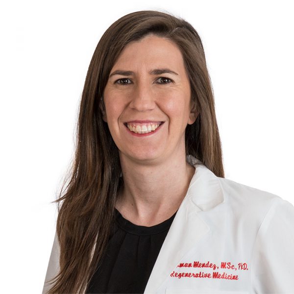 Camila Hochman-Mendez, PhD
Assistant Investigator
Director, Regenerative Medicine Research and Biorepository Core
The Texas Heart Institute
“ Innovations in Cardiovascular Tissue Engineering: Addressing Consistency and Reproducibility”
Abstract: The upcoming presentation on cardiovascular tissue engineering will highlight a suite of innovative solutions addressing the critical challenges of consistency and reproducibility in organ regeneration. Through the development of customized bioreactors with coordinated electromechanical stimulation and automated media changes, there's a marked reduction in contamination risks. Incorporating AI-driven technologies for sterile cell injections and the introduction of an in-situ curable semi-conductive hydrogel further propel the field to new heights. Moreover, the strategic application of cardiac dECM powder significantly advances the maturation of hiPSC-derived cardiomyocytes, optimizing their responsiveness to electromechanical stimuli. With the integration of a cutting-edge magnetic bioreactor system and a cost-effective polylaminin protocol, not only is the quality of cardiomyocyte production enhanced, but also its scalability. These advancements represent pivotal strides in the realm of regenerative medicine, emphasizing the potential to revolutionize organ transplantation processes.
- Biography
-
Dr. Camila Hochman-Mendez serves as the Director of the Regenerative Medicine Research Department and is also the esteemed Director of the College of American Pathologists (CAP)-accredited Texas Heart Institute (THI) Biorepository & Biospecimen Profiling Laboratory. With a rich research portfolio, Dr. Hochman-Mendez's work delves into the intricate interplay of extracellular matrix (ECM) proteins in cell-cell interactions, tissue repair, and regeneration. A particular area of fascination lies in understanding the multifaceted interactions between cells and extracellular molecules and vesicles. At THI, the research lab under Dr. Hochman-Mendez's leadership spearheads innovations in bioartificial organ engineering. Notably, the team's efforts in developing customized bioreactors to accommodate and stimulate "rebuilt" organs have garnered significant attention. Their novel approach utilizing decellularized ECM seeded with iPSC-derived cells offers an exciting ECM-based alternative to traditional organ transplantation. A testament to the lab's pioneering work is the publication detailing a fully revascularized rabbit ventricle repopulated with iPSC-derived cardiomyocytes, endothelial cells, and other cardiac cell types. This significant achievement demonstrated the potential of engineered vessels to maintain patency post-transplantation. With the backing of esteemed institutions like the NIH and AHA, Dr. Hochman-Mendez's current research is setting new benchmarks. The team's exploration into micro-controlled multiparametric bioreactors and avant-garde conductive hydrogels for artificial conduction systems is challenging and redefining existing paradigms in regenerative medicine.
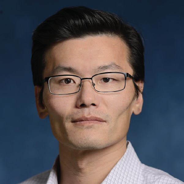 Sean X Sun, Ph.D.
Professor
Department of Mechanical Engineering
Center for Cell Dynamics (CCD)
Institute of NanoBioTechnology
Johns Hopkins University
“Water Dynamics in Cells and Tissues”
Abstract: A large fraction of cell and tissue mass is made of water. The flow of water across the cell surface follows osmotic and hydraulic pressure gradients, and is actively controlled by the cell. This physical fact suggests that the mechanical behavior of cells is intimately connected with cell ionic homeostasis and osmotic control. In this talk, we will describe some of our recent experimental and modeling work on water dynamics in cells. We explore how cells control their cytoplasm water content and therefore the cell size. We will describe how cells can use directional flow of water and ion channels such as the Na+/H+ exchanger to propel themselves and migrate, and in the case of epithelia, pump fluid between tissue compartments. In the later case, epithelial pumping of water can strongly influence tissue shape and morphogenesis. Taken together, we suggest a revision of the kinematic law of biological motion that takes these developments into account.
- Biography
-
Sean Sun is a professor of mechanical engineering and biomedical engineering at Johns Hopkins University and a member of the Institute of NanoBioTechnology (INBT) at JHU. His research has been focused on understanding mechanobiology of the cell using experiments as well as mathematical modeling. In the last ten years, he has developed mechanistic mathematical models to understand cell and nuclear size determination in mammalian and bacterial cells, mammalian cell movement and motility, cytoskeleton and water dynamics in moving cells, fundamental processes behind cellular mechanosensation and force generation. He is especially interested in the interplay between mechanics, forces and biochemistry in cells and tissues, and mechanochemical processes in biology. His lab also has developed simple and robust experimental and imaging methods to measure mechanics of biomolecules and live cells. Prof. Sun is a fellow of the American Physical Society and the American Institute for Medical and Biological Engineering.
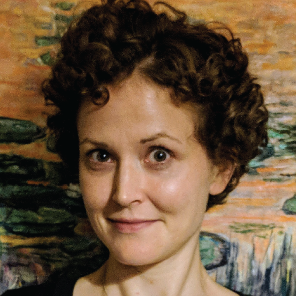 Jeanne Stachowiak, Ph.D.
Professor
T. Brockett Hudson Professorship in Chemical Engineering
Departments of Chemical Engineering & Biomedical Engineering
The University of Texas at Austin
“Intrinsic disorder as an organizing principle for membrane biology ”
Abstract: As the gateway for cellular entry and communication, the surface of the cell holds the answers to critical questions in biology and medicine, while simultaneously providing inspiration for engineered materials and systems. What captivates me about membranes is their ability to precisely and rapidly organize themselves in an environment of staggering complexity. Thousands of distinct protein species reside on cellular membranes, yet functional membrane protein complexes can form within seconds in response to diverse stimuli. Traditionally, structured protein assemblies such as vesicular coats and cytoskeletal filaments have been thought to organize membrane surfaces. In contrast, recent work illustrates that networks composed of proteins with a high degree of intrinsic disorder may provide the necessary flexibility to facilitate efficient assembly of functional protein complexes at membrane surfaces. In particular, our recent work has illustrated that a flexible network of disordered proteins helps to catalyze the assembly of endocytic structures at the plasma membrane. Specifically, we find that an intermediate strength of interaction between these proteins, which leads to liquid-like properties at the macro-scale, maximizes the efficiency of endocytic vesicle assembly. Interestingly, the stability of this catalytic network appears to be controlled by the degree of ubiquitination at endocytic sites, providing a potential mechanism for sensing the presence of transmembrane cargo proteins, many of which are ubiquitinated prior to removal from the plasma membrane. More broadly, the idea of a transition between a stochastic, disordered protein network toward a more ordered structure that is capable of deterministic outcomes provides a template for understanding many processes in membrane biology. At present our group is focused on characterizing these “disorder to order” transitions using a diverse toolset, which includes in vitro biochemistry, quantitative imaging of live engineered cells, and collaborations with theorists who work across a range of length scales. Specific areas of our ongoing work include the role of disordered protein networks in organizing the cytoskeleton, and transmembrane coupling of protein condensates as a novel mechanism of information transfer across biological interfaces.
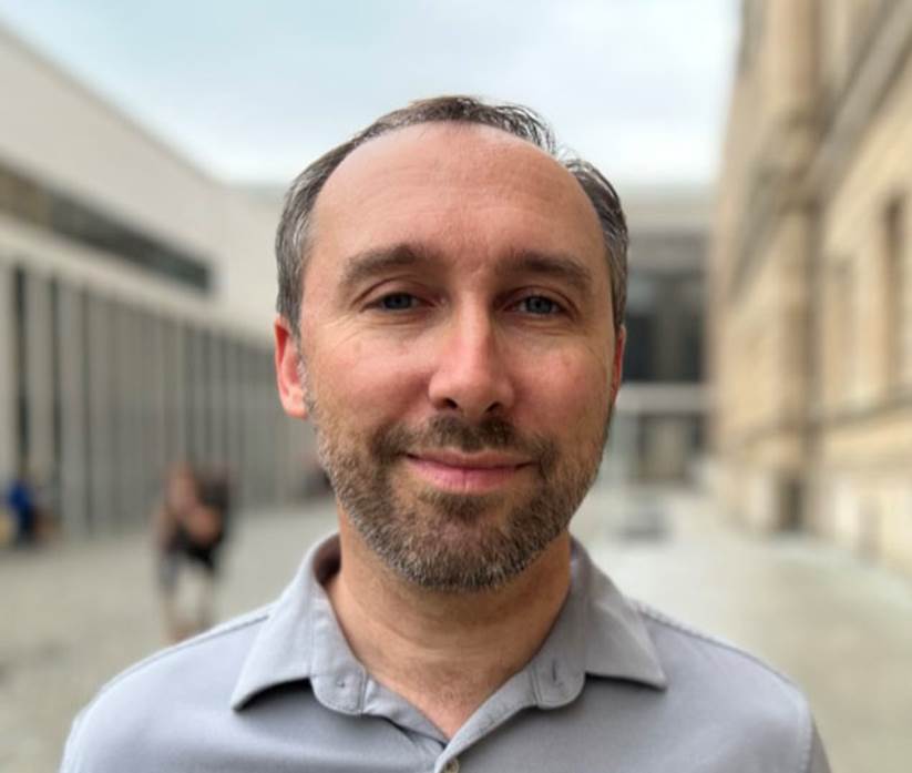 Hernan Garcia, Ph.D.
Associate Professor of Genetics, Genomics and Development
Department of Molecular & Cell Biology and Department of Physics
University of California at Berkeley
“From SHG Microscopy to Space‐Time Optical Fields: Highlights from the PROBE Lab at Brown University”
Abstract: Over the last few decades we have largely identified the repressors and activators that shape gene expression patterns in developing embryos and that, in turn, dictate cellular fates. Yet, despite amassing this great reservoir of knowledge, we are still incapable of predicting how the number, placement and affinity of binding sites for these transcription factors in regulatory DNA dictate gene expression patterns in space and time. Achieving such predictive understanding calls for going beyond molecular parts lists and for obtaining the in vivo biochemical information necessary for fueling theoretical models of transcriptional regulation in developing animals.
In this talk, I will show how we are using physics as a “microscope” to uncover the molecular mechanisms by which activators and repressors dictate transcription in space and time in developing animals. Specifically, using novel quantitative tools that we have developed for precision measurements, I will show that most developmental genes are transcribed in stochastic bursts, and that many transcription factors regulate gene expression by modulating the frequency, duration, and/or amplitude of these bursts. We will then engage in an iterative dialogue between theoretical models and quantitative experiments aimed at revealing the mechanisms underlying this control of transcriptional bursting. Our results challenge the textbook picture of activator and repressor action based on stable protein-protein interactions and call for a description of transcriptional control that acknowledges that the nucleus is not a bag of well-mixed transcription factors. Most importantly, our work sets a path forward for reaching a predictive understanding of cellular decision making and demonstrates how a quantitative dialogue between theory and experiment can shed light on biological mechanisms beyond the reach of even the best super resolution microscopes.
- Biography
-
Hernan G. Garcia is an Associate Professor in the Departments of Molecular & Cell Biology and of Physics at UC Berkeley. As a Physical Biologist, his research aims to uncover the quantitative and predictive principles dictating biological phenomena, with particular emphasis on embryonic development. Hernan is a co-author of the textbook Physical Biology of the Cell and has directed several courses at institutions such as the Kavli Institute for Theoretial Physics at UC Santa Barbara, and the Marine Biological Laboratory in Woods Hole, MA.
 Kimani C. Toussaint, Jr., Ph.D.
Thomas J. Watson, Sr. Professor of Science
Senior Associate Dean for Research and Strategic Initiatives, School of Engineering
Director, Brown-Lifespan Center for Digital Health
Brown University
“From SHG Microscopy to Space‐Time Optical Fields: Highlights from the PROBE Lab at Brown University”
Abstract: The remarkable versatility of optical microscopy has helped to both elucidate our fundamental understanding of biological systems and enable the manipulation of matter at the nanoscale. This talk will highlight the major projects pursued by the laboratory for Photonics Research of Bio/nano Environments (PROBE). Specifically, we will review some of the quantitative secondharmonic generation (SHG) microscopy techniques pursued by the PROBE lab for assessment of a variety of collagenous tissues, including recent work where we consider collagen fibers as fluidlike. We will also discuss an area of recent interest developing and utilizing space‐time optical fields for diffraction‐free propagation and speckle‐resistance in the presence of turbid specimens. Finally, we will briefly discuss our interests in developing optical‐based physiological sensors that work equitably across skin tones.
- Biography
-
K. C. Toussaint, Jr., Ph.D. is the Thomas J. Watson, Sr. Professor of Science, and a Professor and Senior Associate Dean for Research and Strategic Initiatives in the School of Engineering at Brown University. Prior to joining Brown in 2019, Dr. Toussaint was faculty at the University of Illinois at Urbana‐Champaign for 12 years. Dr. Toussaint directs the laboratory for Photonics Research of Bio/nano Environments (PROBE Lab), an interdisciplinary research group working in the areas of quantitative nonlinear optical imaging techniques, structured light, nano‐optics, and optical health‐monitoring techniques that mitigate bias. He is a recipient of a 2010 NSF CAREER Award, the 2014‐2015 Dr. Martin Luther King, Jr. Visiting Associate Professor at MIT, the 2015 Illinois Dean’s Award for Excellence in Research, the 2017 Illinois Everitt Award for Teaching Excellence, and the 2019 Distinguished Promotion Award. Dr. Toussaint is also a Fellow of Optica, SPIE, and AIMBE, as well as a Senior Member in the IEEE. In addition, he is a member of the Board of Reviewing Editors for Science, and has served as a Guest Editor for PNAS. Dr. Toussaint’s work on equitable health technologies has appeared in popular press, including NPR, Politico, CNN, STAT+, and the Boston Globe.
 Gunjan Agarwal, Ph.D.
Professor
Department of Mechanical and Aerospace Engineering
Ohio State University
“A DDR1-Collagen Story in Vascular Biology”
Abstract: Collagen type 1 is the most abundant extracellular matrix protein in adult tissues. The interaction of cells with collagen occurs via specific receptors and regulates both cell and matrix function. We describe here a two-way cross-talk between collagen and the collagen receptor discoidin domain receptor (DDR1). DDR1 is a receptor tyrosine kinases expressed in a variety of mammalian cells. On one hand DDR1 regulates the collagen fibril structure while on the other hand the fibrillar state of collagen impacts receptor function. An altered collagen fibril structure in turn can affect cell-matrix interactions, which have multiple manifestations in health and disease. We elucidate such an effect in vascular biology, wherein the DDR1 knockout mice exhibited enhanced platelet-collagen adhesion and increased thrombogeneity of the vessel wall. Our investigations also reveal that structural alterations in collagen fibril (accompanied by DDR1 activation) exist in vascular diseases such as aortic aneurysms, which can impact platelet adhesion, vascular calcification and mechanics.
- Biography1
-
Dr. Gunjan Agarwal is a Professor in the Department of Mechanical and Aerospace Engineering at the Ohio State University. She received her PhD in Biophysics from the Tata Institute of Fundamental Research in Mumbai, India and came to the US for her post-doctoral training at the Albert Einstein College of Medicine (Bronx, NY) and at Procter and Gamble Pharmaceuticals (Cincinnati OH). After a brief period as a scientist at the Wright Patterson Air Force Base, she joined the Ohio State University in 2003.
Prof. Agarwal’s research interests lie “outside the cell” on extracellular matrix remodeling, with a particular focus on the collagen receptors discoidin domain receptors (DDRs). Like integrins DDRs are ubiquitously expressed and are understood to be important for several diseases such as cancers and fibrosis. Dr. Agarwal has been a pioneer in the field of DDRs and her laboratory has yielded seminal work on collagen fibrillogenesis and cell-matrix interactions. Her ongoing research is directed to unravel the causes and consequences of an altered collagen fibril structure with a particular emphasis in vascular and bone diseases.
Prof Agarwal extensively employs atomic force microscopy (AFM) and other microscopy approaches for her research. She directs a multi user AFM core facility and is the co-director of the interdisciplinary Biophysics graduate program at the Ohio State University. She has published 4 book chapters and over 55 journal articles in well-reputed journals like Acta Biomaterialia, J. Mol. Biol, Small etc. Her research has been continuously funded by the NSF, NIH and the American Heart Association. Dr. Agarwal has also been recognized for her efforts in diversity and inclusion.
 Francesca Taraballi, Ph.D.
Assistant Professor of Orthopedic Surgery
Director, Center for Musculoskeletal Regeneration
Houston Methodist
“Immunoengineering solutions for Translational Medicine”
Abstract: The application of immune engineering in translational medicine has emerged as a transformative approach with the potential to revolutionize disease treatment and personalized healthcare. This interdisciplinary field merges principles of immunology, engineering, and nanotechnology to develop innovative strategies for manipulating the immune system to combat a wide range of diseases. Immune engineering has shown remarkable promise in targeted drug delivery, immunomodulation, and regenerative medicine. By harnessing nanoscale materials and biomaterials, immune engineering enables precise interactions with immune cells and immune-related processes, offering unprecedented opportunities for therapeutic interventions. In this seminar, Dr. Taraballi will present her recent advancements in immune engineering research and its impact on translational medicine application in cancer, regeneration, and infection diseases. She will discuss how nanocarriers and immunomodulatory biomaterials are being designed and tailored to effectively deliver therapeutics to specific disease sites while minimizing off-target effects. Moreover, she will explore how immune engineering is facilitating the development of personalized immunotherapies for cancer and autoimmune disorders. Translational studies focusing on preclinical and clinical trials will be also discussed, highlighting the potential of immune engineering to bridge the gap between bench research and bedside applications. As immune engineering continues to advance, it holds immense promise for reshaping the landscape of translational medicine, ultimately improving patient outcomes, and fostering a new era of precision healthcare.
- Biography
-
Dr. Francesca Taraballi is an assistant professor in Orthopedic Surgery at Houston Methodist Hospital and serves as Director for the Center for musculoskeletal Regeneration affiliated to the Orthopedics and Sports Medicine Department. She is also a faculty member of the Center for RNA therapeutics and have adjuctn position at the Department of Nanomedicine and Neal Cancer Center of the Houston Methodist Hospital. Dr. Taraballi own a master in biochemistry and a Ph.D. in Nanotechnology from the University of Milan- Bicocca.
Dr. Francesca Taraballi works in the field of immune engineering. Her expertise lies in the intersection of nanotechnology, biomaterials, and immunology, where she has made significant contributions to the development of innovative approaches for immune modulation and targeted therapeutics. In her research on immune engineering, Dr. Taraballi focuses on designing and engineering nanoscale materials and platforms that can interact with the immune system in precise and controlled ways. These engineered materials are tailored to either enhance the body's natural immune response or to modulate the immune system to target specific diseases.
One of the key areas of her work involves developing nanocarriers for targeted drug delivery. These nanocarriers are designed to encapsulate therapeutic agents, such as drugs or genetic materials, and deliver them specifically to the desired site of action within the body. By leveraging the immune system's ability to recognize and interact with nanoparticles, Dr. Taraballi's research has resulted in the development of more effective and less toxic therapies for a variety of diseases, including cancer, autoimmune disorders, and infectious diseases. Additionally, she has been actively involved in research related to immunomodulatory biomaterials. These biomaterials are engineered to interact with immune cells and promote specific immune responses, such as promoting tissue repair and regeneration or suppressing excessive inflammation. Such advancements have the potential to revolutionize tissue engineering, regenerative medicine, and immunotherapies. Beyond her scientific contributions, Dr. Taraballi has been an advocate for interdisciplinary collaboration. Her work often involves collaborating with immunologists, clinicians, and materials scientists, fostering a holistic approach to tackle complex biomedical challenges. Overall, Francesca Taraballi's work in immune engineering has significantly advanced the field and holds immense promise for the development of more targeted and personalized approaches to healthcare. Her dedication to harnessing the power of the immune system through nanotechnology has the potential to improve the lives of countless individuals affected by various diseases and conditions.
 Viola Vogel, Ph.D.
Professor of Applied Mechanobiology
Department of Health Sciences and Technology
ETH Zürich, Switzerland
“Mechanoregulation of Cell Niches in Health and Disease”
Abstract: Organ-specific cell niches are not composed of cell monocultures, but of a large variety of cell types which are in close contact to each other and communicate via biochemical and physical factors. Much progress has been made in the molecular understanding of how physical stimuli are sensed and transduced by cells into biochemical signals (mechano-signaling and mechano-transduction), which then regulate gene transcription processes and subsequently cell decision making. Beyond the physical properties of cellular microenvironments, that engineers could easily produce and tune, the reciprocal mechanical signaling between cells and their extracellular environments also involves the stretching of proteins and thus an off-on, or on-off switching of exposed molecular binding sites. Protein stretching can actually switch the structure-function relationships of extracellular as well as of intracellular proteins, which defines the underpinning principles of mechanobiology. Translating what has been learned in mechanobiology (mostly on single cells) to real organs, and finally to the clinic, is hampered by at least two challenges: the lack of nanoscale sensors to probe forces or tissue fiber tensions in healthy versus diseased organs. To bring our knowledge to the patient, we developed a nanosensor to visualize the tensional states of ECM fibers in animal models and in human tissues, with many unexpected findings that were not anticipated from 2D cell culture studies. Underpinning mechano-regulated mechanisms and the significance of these findings in cancer will be discussed.
- Biography
-
Viola Vogel’s research in Bioengineering focuses on how to exploit emerging knowledge in Mechanobiology for Applications in Tissue Engineering, Regenerative Medicine and to treat inflammatory diseases. With experimental and computational tools, she discovered various structural mechanisms how cells sense mechanical forces and convert them into biochemical signals, ultimately altering molecular transcription processes. She graduated with a PhD in Physics from the University of Frankfurt (1987) with research conducted at the Max-Planck Institute for Biophysical Chemistry in Göttingen, for which she received the Otto- Hahn Medal. After her postdoctoral studies in Physics at UC Berkeley, she started her academic career at the University of Washington Seattle in Bioengineering (1990-2004), where she was the founding Director of the Center for Nanotechnology (1997-2003). When moving to ETH Zurich in 2004, she initially joined the Department of Materials and received an ERC Advanced Grant (2009). She then co-founded the Department of Health Sciences and Technology (2012) and chaired the Department from 2018-2020. She is currently Einstein Fellow at the Charité Berlin and Elected Member of the Leopoldina, the Berlin-Brandenburg Academy of Sciences, and of both the National Academy of Engineering (NAE) and the National Academy of Sciences (NAS). She received an Honorary Doctor of Philosophy from Tampere University, Finland 2012.
 Shreya Raghavan, Ph.D.
Assistant Professor, Biomedical Engineering
Texas A&M University
Adjunct Assistant Professor, Nanomedicine
Houston Methodist Research Institute
“Metastatic cancer microenvironments: intersecting roles of biomaterials, immunobiology and mechanics”
Abstract: A majority of primary tumors are detectable early, removable via surgery, or treatable with chemotherapy. However, tumor recurrence, metastasis, and chemoresistance are significant causes of mortality in many tumor types. Like any other organ in the body, tumors harbor cancer stem cells, capable of regenerating the tumor or allowing it to escape a chemotherapy insult. Cancer stem cells are also responsible for aggressive metastasis and chemoresistance. Typical to organogenesis and regeneration, tumors actively recruit supporting stromal cells, bypass immune targeting, and recur and metastasize by harnessing microenvironmental cues. Within this context, how cancer stem cells evade immune suppression and metastasize is of significant interest. Unpacking this interaction will lead to the development of new targeted and immunotherapies to curb metastatic spread and its associated mortality. Here, we will look through a biomedical engineer’s lens to identify variables within the cancer stem cell/immune axis, construct engineered models to isolate the variable and further our understanding of how cancers metastasize.
- Biography
-
Dr. Raghavan is an Assistant Professor in the Department of Biomedical Engineering at Texas A&M University. She has a PhD in Biomedical Engineering from Wake Forest University/Virginia Tech, and was a Ruth L. Kirchstein National Research Service Postdoctoral Fellow in Cancer Tissue Engineering at the University of Michigan. At A&M, Dr. Raghavan directs the Stem Cell, Cancer and Immune Tissue Engineering lab, where she builds engineered micro-tissues and micro- environments to study how cancers grow and disseminate. Her approaches integrate mechanobiology, biomaterials and microenvironment engineering to ask questions that intersect the cancer stem cell/immune axis. Her work is funded by the NIH/NCI through an R37 MERIT award, the Department of Defense, and the state cancer agency, Cancer Prevention and Research Institute of Texas. She is also an award winning teacher, recognized for her inclusive pedagogy in the undergraduate classroom by a Montague Scholars Award from Texas A&M University. Dr. Raghavan is an advocate and ally for justice and equity, diversity and inclusion and works actively towards dismantling systemic processes that hold academics behind in STEM. She serves on the DEI Committee of the Society for Biomaterials, to uphold the values of the society to promote equity for all its members.
 Audrey K. Bowden, Ph.D.
Dorothy J. Wingfield Phillips Chancellor Faculty Fellow
Faculty Head, Nicholas S. Zeppos College
Associate Professor, Biomedical Engineering
Associate Professor, Electrical and Computer Engineering
Affiliate Faculty, Vanderbilt Institute of Global Health
Affiliate Faculty, Vanderbilt Institute for Surgery and Engineering
“New Diagnostic Tools to Enhance the Sensitivity and Specificity of Detection for Early-Stage Bladder Cancer”
Abstract: Bladder cancer (BC) — the 4th most common cancer in men and the most expensive cancer to treat over a patient’s lifetime — is a lifelong burden to BC patients and a significant economic burden to the U.S. healthcare system. The high cost of BC stems largely from its high recurrence rate (>50%); hence, BC management involves frequent surveillance. Unfortunately, the current in-office standard-of-care tool for BC surveillance, white light cystoscopy (WLC), is limited by low sensitivity and specificity for carcinoma in situ (CIS), a high-grade carcinoma with high potential to metastasize. Early detection and complete eradication of CIS are critical to improve treatment outcomes and to minimize recurrence. The most promising macroscopic technique to improve sensitivity to CIS detection, blue light cystoscopy (BLC), is costly, time-intensive, has low availability and a high false-positive rate. Given the limitations of WLC, we aim to change the paradigm around how BC surveillance is performed by validating new tools with high sensitivity and specificity for CIS that are appropriate for in-office use. In this seminar, I discuss our innovative solutions to improve visualization and mapping of suspicious lesions as well as to create miniature tools for optical detection based on optical coherence tomography (OCT). OCT and its functional variant, polarization-sensitive OCT, can detect early-stage BC with better sensitivity and specificity than WLC. We discuss the critical technical innovations necessary to make OCT and PS-OCT a practical tool for in-office use, and new results from recent explorations of human bladder samples that speak to the promise of this approach to change the management of patient care.
- Biography
-
Audrey K Bowden is the Dorothy J. Wingfield Phillips Chancellor Faculty Fellow and Associate Professor of Biomedical Engineering (BME) and of Electrical and Computer Engineering (ECE) at Vanderbilt University. Prior to this, she served as Assistant and later Associate Professor of Electrical Engineering and Bioengineering at Stanford University. Dr. Bowden received her BSE in Electrical Engineering from Princeton University, her PhD in BME from Duke University and completed her postdoctoral training in Chemistry and Chemical Biology at Harvard University. She is a Fellow of SPIE, a Fellow of AIMBE, a Fellow of Optica and a recipient of numerous awards. She currently serves on the Board of Directors of SPIE. Her research interests include biomedical optics (particularly optical coherence tomography and near infrared spectroscopy), microfluidics, and point-of-care diagnostics.
Tuesday, February 28 | 4:00 pm
 Sang Woo Seo, Ph.D.
Associate Professor
School of Chemical and Biological Engineering
Seoul National University, South Korea
“Microbial Synthetic Biology”
Abstract: The growing interest in the bio-based industry as an alternative production route has given rise to numerous efforts to redesign existing biological systems or synthesize new ones. Imagined to build a system, we require various molecular parts/tools and also need insights on assembling the subset or entire biological systems to exhibit specific functions. The former can be achieved by “Synthetic Biology” approach (bottom-up), and the latter can be achieved by “Systems Biology” approach (top-down). Coordinating these two approaches is mandatory to achieve goals effectively. In this talk, I will describe recent efforts to develop synthetic biology tools such as selection-free tunable plasmid (STAPL) system, synthetic protein quality control (ProQC) system, and biosensor-based adaptive laboratory evolution (ALE) system. Furthermore, their applications in designing and optimizing synthetic cell factories, including newly isolated fast-growing Vibrio sp. dhg to convert marine biomass will be described.
- Biography
-
SangWoo Seo is an Associate Professor in the School of Chemical and Biological Engineering at Seoul National University (SNU), South Korea. He earned a B.S. in Chemical Engineering from POSTECH (Pohang University of Science and Technology) in 2007 and a Ph.D. in Chemical Engineering from POSTECH in 2012, where he worked with Prof. Gyoo Yeol Jung to develop synthetic biology tools in E. coli. He was a postdoctoral scholar in the Department of Bioengineering at UCSD (Prof. Bernhard Palsson’s lab), where he applied multi-omics analyses to reconstruct transcriptional regulatory networks in E. coli. At SNU, his research group focuses on i) elucidation of complex regulatory networks in microorganisms by using next-generation sequencing (NGS) based multi-omics analyses, ii) development of novel synthetic biology tools, and iii) redesigning microorganisms for the production of chemicals, fuels, food additives, pharmaceuticals, and therapeutics, for environmental bioremediation, and for biomedical applications. Several microorganisms such as E. coli (including Nissle 1917), Vibrio natriegens, Corynebacterium glutamicum, Bacillus megaterium, Methanotrophs, Lactobacillus, Saccharomyces cerevisiae, Yarrowia lipolytica, and Pichia pastoris are used as host systems.
Tuesday, March 7 | 4:00 pm
 Arvind P. Pathak, Ph.D.
Professor
Depts. of Radiology, Biomedical and Electrical Engineering
The Sidney Kimmel Comprehensive Cancer Center
Institute for NanoBioTechnology
Institute for Computational Medicine
The Johns Hopkins University School of Medicine
““IMAGE-BASED” SYSTEMS BIOLOGY - The Next Frontier in Bioengineering”
- Biography
-
Dr. Pathak (www.pathaklab.org) is a biomedical engineer, ideator and mentor whose mission is to “improve lives via the transformative power of images”. He received a BS in Electronics Engineering from the University of Poona, India. He received his PhD from the joint program in Functional Imaging between the Medical College of Wisconsin and Marquette University, where he was a Whitaker Foundation Fellow. He completed a postdoctoral fellowship in Molecular Imaging in the Dept. of Radiology at the Johns Hopkins University School of Medicine. At Johns Hopkins University (JHU), Dr. Pathak is Professor in the Schools of Medicine (Radiology and Oncology) and Engineering (Biomedical and Electrical Engineering). He is also a member of the Sidney Kimmel Comprehensive Cancer Center, the Institute for NanoBioTechnology (INBT), and the Institute for Computational Medicine (ICM). As globally recognized expert on functional and molecular imaging, his research has been recognized by multiple awards such as the Bill Negendank Award from the International Society for Magnetic Resonance in Medicine (ISMRM) given to “outstanding young investigators in cancer MRI” and the Career Catalyst Award from the Susan Komen Breast Cancer Foundation. Dr. Pathak has served in multiple leadership roles spearheading imaging initiatives, mentoring junior faculty, review panels for national and international funding agencies, and journal editorial boards. He was the Chair of the Cancer Section for the International Society for Magnetic Resonance in Medicine, serves on the Executive Council of the Microcirculatory Society (MCS) and was the Chair of the NIH’s Emerging Imaging Technologies in Neuroscience (EITN) study section. He directs an interdisciplinary, nurturing and continuously funded laboratory, and has mentored almost a 100 award- winning (e.g. Fulbright and Howard Hughes Fellows) students. He has served on multiple diversity initiatives and is dedicated to mentoring the next generation of thought leaders in bioengineering. He was the recipient of the ISMRM’s Outstanding Teacher Award, was a JHU Career Champion nominee, and recognized as a 125 Hopkins Hero by the School of Medicine for his “outstanding dedication to the core values of Johns Hopkins University”.
Tuesday, February 21 | 4:00 pm
 Juliane Nguyen, Ph.D.
Division of Pharmacoengineering and Molecular Pharmaceutics
Eshelman School of Pharmacy
University of North Carolina at Chapel Hill
“Engineering Non-Invasive Drug Depots and Auxetic Patches for Cardiac Applications and Dynamic Organ Repair”
Abstract: Dr. Nguyen’s talk introduces an advanced approach to creating non-invasive therapeutic depots that use zipper-mediated crosslinking. She discusses its use in engineering frameworks for creating scaffold-free delivery methods for cardiac repair. Unlike conventional drug depots, this new generation of drug depots can be administered and removed non-invasively. Thus, they do not require surgery. Furthermore, Dr. Nguyen will discuss the development of auxetic patches for dynamic organ repair. Current patches do not conform to the complex mechanics of dynamic organs, such as the lung or heart. To address these limitations, Dr. Nguyen’s lab has developed a scalable patch platform that is instantly adhesive and able to conform to volumetric changes for dynamic organ and wound repair.
- Biography
-
Juliane Nguyen, Ph.D., is an Associate Professor and Vice Chair in the Division of Pharmacoengineering and Molecular Pharmaceutics, School of Pharmacy, at the University of North Carolina at Chapel Hill. Her lab develops complex biologics for treating myocardial infarction, cancer, colitis, and other diseases. Dr. Nguyen’s work has been recognized with the NYSTAR faculty award, the NSF CAREER Award (2018), the Young Innovator Award from the Biomedical Engineering Society (2019), and the AAPS Emerging Leader Award (2019). She received her Ph.D. in Pharmaceutical Sciences from the Philipps-University of Marburg (Germany), where she was mentored by Dr. Thomas Kissel. She then trained at UCSF under Dr. Frank Szoka, where she was a Deutsche Forschungsgemeinschaft Postdoctoral Fellow. Dr. Nguyen is currently a standing member of the NIH Drug and Biologic Therapeutic Delivery (DBTD) study section. She is also the Executive Editor of Advanced Drug Delivery Reviews.
Tuesday, February 7 | 4:00 pm
 Edward Botchwey, Ph.D.
Professor
The Wallace H. Coulter Department of Biomedical Engineering
Georgia Institute of Technology
“Unlocking the Power of Bioactive Lipid Signaling for Immunoengineering and Regenerative Medicine”
Abstract: Innate immune cells play a crucial role in tissue growth, stability, and injury recovery. They guide blood vessel restructuring, activate local stem and progenitor cells, and direct tissue repair, such as in muscle and skin. To leverage this innate ability, immunoregenerative biomaterials are being developed that target specific subpopulations of monocytes/macrophages to promote repair by controlling their recruitment, positioning, differentiation, and function in damaged tissues. Monocyte and macrophage phenotypes range from inflammatory (M1) to pro-regenerative (M2) and their functions are heavily influenced by the injury environment. It's believed that dividing labor among different subpopulations of monocytes and macrophages could optimize regenerative functions over inflammatory ones. However, the delicate balance between necessary inflammatory and regenerative functions of myeloid cells is not yet fully understood. This talk will examine distinct populations of monocytes and macrophages, their role in various injury environments, and smart biomaterial design strategies that harness the innate inflammatory response to improve healing.
- Biography
-
Edward Botchwey received a B.S. degree in Mathematics from the University of Maryland at College Park in 1993 and both M.E. and Ph.D. degrees in Materials Science Engineering and Bioengineering from the University of Pennsylvania in 1998 and 2002, respectively. He is currently a Professor in the Wallace H. Coulter Department of Biomedical Engineering at Georgia Tech and Emory University. Dr. Botchwey is a former Ph.D. fellow of the National GEM Consortium, a former postdoctoral fellow of the UNCF-Merk Science Initiative, and a recipient of the Presidential Early Career Awards for Scientists and Engineers from the National Institutes of Health and the Society for Biomaterials Mid-Career Award. Dr. Botchwey’s research focuses on how transient control of immune response using bioactive lipids can be exploited to control the trafficking of stem cells, enhance tissue vascularization, and resolve inflammation.
Tuesday, January 24 | 4:00 pm
 Horacio Espinosa, Ph.D.
Northwestern University
“Micro and Nano Technologies for Cellular Engineering and Material Discovery”
Abstract: The ability to control fluidic, electrical, and mechanical fields using micro and nanotechnologies is enabling key advances in cellular engineering and material discovery. In this presentation, I will address two examples: i) the role of microfluidic platforms in genome engineering and temporal cell analysis, and ii) the use of microsystems for in-situ electron microscopy characterization of low dimensional materials. In cellular engineering, a critical step is the intracellular delivery of gene editing machinery and subsequent nondestructive, temporal analysis of cells. Both can be achieved by cell membrane permeabilization, as part of workflows employed in the investigation of molecular mechanisms of disease, pharmacological screening, and development of new therapeutics. Specifically, I will discuss several microfluidic platforms created in my lab, ranging from a fully automated nanofountain probe electroporation (NFP-E) system, which provides single- cell manipulation with superior cell viability and efficiency, and a live cell analysis device (LCAD) for nondestructive and temporal cellular analysis. A critical aspect of the technology is the possibility of perturbing cell state and downstream pathways. I will present single cell RNA sequencing data analysis to show that microfluidic technology leads to significantly reduced cell stress response and much higher control of molecular payload when compared to standard delivery methods. In a second example, I will discuss microsystems to investigate size scale effects on the mechanics of 1D and 2D materials. These materials are being employed in the development of next- generation electronics, optical, and sensor technologies, as well as in energy production and storage techniques, e.g., supercapacitors, solar cells, and battery electrodes. Such applications involve frequent mechanical deformations such as stretching and bending, so the lifespan (integrity and reliability) of the material is a critical feature. In this context, I will present in situ electron microscopy experiments and atomistic models we have used to understand deformation and failure modes in metallic nanowires and transition metal dichalcogenides. Implications on the development of atomistic models with predictive capabilities, in the spirit of the materials genome initiative, will be highlighted.
- Biography
-
Horacio D. Espinosa is the James and Nancy Farley Professor of Manufacturing and Entrepreneurship, Professor of Mechanical Engineering, and the Director of the Theoretical and Applied Mechanics Program at the McCormick School of Engineering, Northwestern University. He received his Ph.D. in Solid Mechanics from Brown University in 1992. Espinosa has made contributions in the areas of deformation and failure of materials, design of micro- and nano- systems, in situ microscopy characterization of nanomaterials, and microfluidics for single cell manipulation and analysis. He has published over 300 technical papers on these topics. Espinosa received several awards including the Prager Medal from the Society of Engineering Science, the Society for Experimental Mechanics Murray and Sia Nemat Nasser Medals, and the ASME Drucker and Thurston awards. He is a member of the National Academy of Engineering (NAE), foreign member of Academia Europaea, the European Academy of Arts and Sciences, the Russian Academy of Engineering, and Fellow of AAAS, ASME, SEM, and AAM. He was the President of the Society of Engineering Science in 2012 and is a member of the IUTAM General Assembly.
Tuesday, January 10 | 4:00 pm
 Amanda Nash
Ph.D. Candidate
Bioengineering Department
Rice University
“Development of implantable cytokine factories for controlled modulation of the immune response”
Abstract: Pro-inflammatory cytokines have been approved by the FDA for the treatment of metastatic melanoma and renal carcinoma. However, effective cytokine therapy is limited by a short half-life in circulation and the severe adverse effects associated with high systemic exposure. To overcome these limitations, we developed a cytokine delivery platform composed of polymer encapsulated epithelial cells that produce natural cytokines. Local administration of these cytokine factories demonstrated predictable dose modulation and provided effective cancer immunotherapy in multiple preclinical models without systemic toxicities. These preclinical models include ovarian cancer, pancreatic cancer, melanoma, and colorectal cancer. Interestingly, we found that the local concentration (IP space) was greater than 100x higher than the systemic concentration (blood), demonstrating the ability of the platform to deliver cell- generated cytokines in vivo and create a high local concentration of cytokines with limited peripheral exposure. Further, we have demonstrated the ability to readminister these cytokine factories as well as engineer them with a safety switch which provides flexibility with dosing and administration in the clinic. Our data confirmed local increases in the activation (CD25+CD8+) and proliferation (Ki67+CD8+) of cytotoxic T cells within the IP space of cytokine factory treated mice. Significantly, this platform also produced local and systemic T cell biomarker profiles that predict efficacy without toxicity in non-human primates. Overall, our findings demonstrate the safety and efficacy of cytokine factories in preclinical animal models and have been translated into clinical testing for the treatment of ovarian cancer in humans.
Special Seminar
Tuesday, November 29 | 3:00 pm
 Michael R. King, Ph.D.
J. Lawrence Wilson Professor and Department Chair of Biomedical Engineering
Vanderbilt University
“The shear force is all around us: Mechanotransduction of cancer and immune cells in fluid flow”
Abstract: Many types of cancer metastasize via the bloodstream, where circulating tumor cells (CTCs) originating from the primary tumor can travel through the circulation or lymphatic system and engraft in distant organs. Previously our laboratory found that cancer cells exposed to physiological levels of fluid shear stress (FSS) are dramatically more susceptible to undergoing apoptosis via TRAIL protein, inspiring a new therapeutic drug delivery approach to target metastatic cells in the circulation. The FSS response of CTCs and their neutralization by lipid nanoparticle conjugation to the surface of circulating immune cells has been demonstrated with in vitro cell line experiments, orthotopic mouse models of metastasis, and analysis of primary CTC aggregates isolated from metastatic cancer patients. We later learned that this shear stress is primarily mediated by Piezo1 activation, and is modulated by interacting with aggregated stromal cells such as cancer-associated fibroblasts. Interestingly, we recently found that FSS activation of Piezo1 also dramatically enhances the activation of T cells and dendritic cells, which may have important implications for various immunotherapy applications.
Special Seminar
Thursday, December 01 | 3:00 pm
 Cynthia A. Reinhart-King, PhD
University Distinguished Professor
Cornelius Vanderbilt Professor
Biomedical Engineering Senior Associate Dean for Research
School of Engineering
Vanderbilt University
“The intersection of mechanobiology and cellular metabolism in cancer”
Abstract: During solid tumor progression, cells undergo mechanical and metabolic changes that help to fuel metastasis. To move, cells must utilize ATP to fuel the the cellular contractility and forces that sustain migration, however very little is known about how the metabolic state of a cell affects its ability to migrate and vice versa. In this talk, I will describe my lab’s efforts to understand the forces driving cell movements in the tumor microenvironment and the energy required for movement. Combining tissue engineering approaches, mouse models, and patient samples, we create and validate in vitro systems to understand how cells navigate the tumor stroma environment with the goal of identifying novel targets of cancer metastasis. Microfabrication and native biomaterials are used to build mimics of the paths created and taken by cells during metastasis. Using these platforms, we have described a role for a balance between cellular energetics, cell and matrix stiffness, and confinement in determining migration behavior. Moreover, we have extended this work into investigating the intersection of the diabetes and the diabetic tissue microenvironment with tumor progression, showing that mechanical changes in the tissue due to diabetes can promote cancer. Overall, our work has demonstrated key mechanical drivers of metastasis within the tissue microenvironment.
Tuesday, November 08 | 4:00 pm
 Kristi Kiick, Ph.D.
Professor and Chair,
Department of Biomedical Engineering
Blue and Gold Distinguished Professor
Department of Materials Science and Engineering
University of Delaware
“Engineering bioelastomers for applications in treating human disease”
Abstract: Among many types of biomaterials, micro- and nano-scale structures offer important approaches for mediating biological events, in large part because of their ability to effectively target specific tissues and cells. The Kiick group has employed a combination of biosynthetic tools, bioconjugation strategies, and biomimetic assembly to produce elastomeric hydrogels and thermoresponsive peptide nanostructures derived from naturally occurring elastomeric proteins. Because of their composition and assembly, these materials have shown promise for possible applications in treating pathologies of tissues and joints.
- Biography
-
Kristi Kiick is a Professor and Chair of the Department of Biomedical Engineering, and the Blue and Gold Distinguished Professor of Materials Science and Engineering, at the University of Delaware. She also holds affiliated faculty appointments in the Department of Biological Sciences at the University of Delaware and in the School of Pharmacy at the University of Nottingham, where Kiick has conducted research as a Leverhulme Visiting Professor and Fulbright Scholar. Her internationally recognized research focuses on the synthesis, characterization, and application of protein, peptide, and self-assembled materials for applications in tissue engineering, drug delivery, and bioengineering, with specific research in cardiovascular, musculoskeletal, and wound healing approaches.
A Fellow of the National Academy of Inventors, the American Association for the Advancement of Science, and of the American Chemical Society, she has published more than 150 articles, book chapters, and patents, and has delivered over 200 invited and award lectures. Kiick’s honors have included several awards (Camille and Henry Dreyfus Foundation New Faculty, Beckman Young Investigator, NSF CAREER, DuPont Young Professor, and Delaware Biosciences Academic Research Award) as well as induction also as a fellow of the American Institute for Medical and Biological Engineering and of the American Chemical Society Division of Polymer Chemistry. She served as the Deputy Dean of the UD College of Engineering for 8 years, and also serves on the advisory and editorial boards for multiple international journals and research organizations. Kiick received her bachelor of science in chemistry from UD as a Eugene du Pont Memorial Distinguished Scholar, where she graduated summa cum laude, and a masters of science in chemistry as an NSF graduate fellow at the University of Georgia. She worked in industry (Kimberly Clark Corporation) as a research scientist prior to obtaining masters of science and doctoral degrees in polymer science and engineering at the University of Massachusetts Amherst, completing her doctoral research at the California Institute of Technology as a recipient of a National Defense Science and Engineering Graduate (NDSEG) fellowship.
Tuesday, October 25 | 4:00 pm

Brian Applegate, Ph.D.
Professor of Otolaryngology–Head & Neck Surgery and Biomedical Engineering
University of Southern California
“Functional and morphological imaging of the middle and inner ear with Optical Coherence Tomography and Vibrometry”
Abstract: Over the past decade we have been developing Optical Coherence Tomography and Vibrometry (OCTV) to measure the detailed morphology and vibratory response of the ear. With micron scale spatial resolution and subnanometer sensitivity to vibration it is well suited to measuring the spatially resolved vibratory response of both the inner and middle ear. In small animals, it is possible to image directly through the bone of the otic capsule for noninvasive spatially resolved vibrometry of the cochlear partition. In humans as well as small animals, it’s possible to image the tympanic membrane and ossicles through the ear canal to reveal the vibratory response of the middle ear. Nominally, this approach allows for the measure of vibratory response along the light path of the instrument, hence 1-D. In recent work we have developed a system that incorporates 3 separate sample arms in a single interferometer. This allows us to reconstruct the full 3-D vector of motion. We have also begun translating OCTV for use in humans with the development of hand-held devices which can be used in the clinic. The seminar will be split between these two projects, outlining the technical design and discussing recent results for each.
- Biography
-
Dr. Applegate is a Professor of Otolaryngology–Head & Neck Surgery and Biomedical Engineering at the University of Southern California. He received his Ph.D. in physical chemistry from The Ohio State University. He won a National Institutes of Health postdoctoral fellowship grant to continue his training at Duke University in biomedical engineering. Upon completing his fellowship, he joined the faculty of Texas A&M University where he worked for 12 years advancing to the rank of Associate Professor of Biomedical Engineering. He moved to the University of Southern California in 2019 where he joined his long time collaborator to continue their work on functional imaging of the ear. Throughout his career, his research has been supported by grants from the National Science Foundation, including the NSF Career award, the Department of Defense, and the National Institutes of Health. He was elected a fellow of the Optical Society of America in 2016. He has served as an Associate Editor for IEEE Transactions on Medical Imaging and Optics Letters. He is a standing member of the Imaging Guided Interventions and Surgery [IGIS] study section at the National Institutes of Health. His research interests are broadly to develop novel biophotonic technologies and apply them to the diagnosis and monitoring of human disease.
Dr. Applegate’s research interests are broadly to develop novel biophotonic technologies and apply them to the diagnosis and monitoring of human disease. Throughout his career, his research has been supported by grants from the National Science Foundation, including the NSF Career award, the Department of Defense, and the National Institutes of Health. He is a fellow of the Optical Society of America and senior member of SPIE.
Dr. Applegate is a Professor of Otolaryngology–Head & Neck Surgery and Biomedical Engineering at the University of Southern California. His research interests include the development of novel biophotonic technologies with applications in the diagnosis and monitoring of human disease. He has a particular interest in the development and translation of functional imaging technologies. He is a fellow of OSA and senior member of SPIE.
Dr. Applegate is a Professor of Otolaryngology–Head & Neck Surgery and Biomedical Engineering at the University of Southern California. His research interests include the development of novel biophotonic technologies with applications in the diagnosis and monitoring of human disease. He has a particular interest in the development and translation of functional imaging technologies. Throughout his career, his research has been supported by grants from the National Science Foundation, including the NSF Career award, the Department of Defense, and the National Institutes of Health.
Tuesday, October 18 | 4:00 pm
Ahmad (Mo) Khalil
Associate Professor of Biomedical Engineering
Associate Director of the Biological Design Center
Boston University
“Programming at the interface of synthetic and natural cellular networks”
Abstract: Constructing and introducing synthetic biological systems into living cells presents unique opportunities to examine design principles of biological networks and to harness this understanding for creating new therapeutic modalities. In this talk, I will describe two studies that examine and exploit the interface between artificial circuits and the natural pathways with which they interact. In the first study, I will describe how we are using synthetic circuits in yeast to understand how gene regulatory specificity can emerge in eukaryotic transcriptional networks. As we better understand the design rules of transcription circuits, we have also become interested in translating our insights into platforms for creating programmable cellular therapies. The second study focuses on cell signaling systems, where our goal is to design synthetic systems that deliberately interface with and gain the ability to precisely perturb GPCR signaling networks, with implications for basic discovery and new therapeutics.
- Biography
-
Ahmad (Mo) Khalil is the Dorf-Ebner Distinguished Associate Professor of Biomedical Engineering and the Founding Associate Director of the Biological Design Center at Boston University. He is also a Visiting Scholar at the Wyss Institute for Biologically Inspired Engineering at Harvard University, and Co-Director of a NIH/NIGMS T32 PhD Training Program in synthetic biology. His research is broadly interested in learning the design principles underlying the function and evolution of biological networks, and in turn developing methods to predictively engineer them to program therapeutically-useful cellular functions. His lab is also developing methods to recreate and harness the process of evolution, specifically by developing novel technologies, such as the eVOLVER, that enable this process to occur rapidly, autonomously, and at scale in the laboratory. He is recipient of numerous awards, including a Schmidt Science Polymath Award, Presidential Early Career Award for Scientists and Engineers (PECASE), DoD Vannevar Bush Faculty Fellowship, NIH New Innovator Award, NSF CAREER Award, DARPA Young Faculty Award, and Hartwell Foundation Biomedical Research Award, and he has received numerous awards for teaching excellence at both the Department and College levels. Mo was an HHMI Postdoctoral Fellow with Dr. James Collins at Boston University. He obtained his Ph.D. from MIT and his B.S. (Phi Beta Kappa) from Stanford University.
Tuesday, October 04 | 4:00 pm
Moji Karimi, M.S.
CO-Founder and CEO
Cemvita Factory
“Synthetic Biology meets Energy Transition; a Nature-Inspired Future”
Abstract: Energy Transition is upon us and is bringing about a fundamental change in many industries. Some companies are reacting to the change, and some see it as an opportunity to reinvent who they are and create a brighter future (and as shocking as it is, some companies aren’t doing anything at all!). Energy transition has presented the CEOs with a classic innovator’s dilemma. In this talk Moji describes how engineered and scaled synthetic biology can be a solution to decarbonize hard-to-abate sectors while creating new revenue streams in demand for lower-carbon products and realizing the promise of the bio-economy.
Tuesday, September 27 | 4:00 pm

Douglas Lauffenburger, PhD
Department of Biological Engineering
Massachusetts Institute of Technology
“Cross-Species Translation of Biological Information via Computational Systems Modeling Frameworks”
Abstract: A vital challenge that the vast majority of biological research must address is how to translate observations from one physiological context to another—most commonly from experimental animals (e.g., rodents, primates) or technological constructs (e.g., organ-on-chip platforms) to human subjects. This is typically required for understanding human biology because of the strong constraints on measurements and perturbations in human in vivo contexts. Direct translation of observations from experimental animals to human subjects is generally unsatisfactory because of significant differences among organisms at all levels of molecular properties from genome to transcriptome to proteome and so forth. Accordingly, addressing inter-species translation requires an integrated experimental/computational approach for mapping comparable but not identical molecule-to-phenotype relationships. This presentation will describe methods we have developed for a variety of cross-species translation examples, demonstrated on applications in inflammatory and neurological pathologies.
- Biography
-
Douglas Lauffenburger is Ford Professor of Bioengineering in the Departments of Biological Engineering, Chemical Engineering, and Biology at MIT. He was the founding Head of the Department of Biological Engineering at MIT, and served in that capacity 1998-2019. The Lauffenburger research program centers on systems biology approaches to cell-cell communication and cell signaling in pathophysiology, emphasizing translational application to therapeutics discovery and development in cancer, pathogen infection, and inflammatory disease. Lauffenburger has co-authored the monograph Receptors: Models for Binding, Trafficking & Signaling (Oxford Press, 1993) and co-edited the book Systems Biomedicine: Concepts and Perspectives (Elsevier Press, 2010). More than 130 doctoral students and postdoctoral associates have undertaken research education under his supervision. He is a member of the National Academy of Engineering and American Academy of Arts & Sciences, and a fellow of the American Association for Advancement of Science and the American Scientific Affiliation. Lauffenburger has served as President of the Biomedical Engineering Society, Chair of the College of Fellows of American Institute for Medical & Biological Engineering, on the Advisory Council for the National Institute of General Medical Sciences, and as a co-author of the 2009 National Research Council report A New Biology for the 21st Century. He was a co-awardee of the National Academy of Engineering Gordon Prize for Innovation in Engineering & Technology Education in 2021.
Thursday, August 18 | 3:00 pm
Heungsoo Shin, PhD
Professor
Department of Bioengineering
Hanyang University, Korea
“Biomaterials-based delivery of signaling molecules and cells for tissue engineering and regenerative medicine”
Abstract: Tissue engineering and regenerative medicine have gained considerable interest in recent years due to the potential to treat patients with chronic or degenerative diseases. For example, bone fracture associated with increase in patients with osteoporosis and other injuries becomes one of the large clinical problems, which requires tissue engineering strategy because of limited supply of graft materials. Tissue healing is complex processes involving regulation of inflammatory response after injury, migration, tissue-specific induction of uncommitted mesenchymal stem cells, and remodeling of loosely formed pre-mature tissue. Therefore, it is of importance to consider engineering approaches to regulate multi-facet processes of these tissue healing events. Given that, we have been developing biomaterials as carriers of various cell-instructive signals or stem cells to actively guide tissue regeneration. For example, biomaterials with controlled substrate-mediated delivery of proteins, peptide, or small molecules have been developed to enhance osteogenesis of stem cells and suppression of inflammatory response. The combination of stem cells with these materials have been used to engineer multi-cellular composite 3D spheroids for the fabrication of structurally and functionally reliable in vitro artificial 3D tissues. In addition, strategies for encapsulation of spheroids within biocompatible hydrogels have been explored for recapitulation of cellular microenvironment of engineered tissue of the desired shape with enhanced specific biological functions. This presentation will summarize and discuss our endeavor to engineer biomaterials in regard to how we design them enabling delivery of stem cells and signaling molecules, in particular for regeneration of bone tissue.
- Biography
-
Dr. Heungsoo Shin is currently Professor with tenure in the Department of Bioengineering. He received his B.S. and M.S. degree in the Department of Industrial Chemistry from Hanyang University, Korea, and his Ph.D. degree in the Department of Bioengineering from Rice University, USA (2004) from the guidance of Dr. Antonios G. Mikos. He was a postdoctoral researcher at the Department of Mechanical Engineering, Georgia Institute of Technology, USA with Dr. Andres J. Garcia before joining the faculty in the Department of Bioengineering, Hanyang University, Korea in 2006 as assistant professor. His main research areas lie in (1) Development of biomimetic materials for delivery of bioactive molecules and stem cells, (2) Surface modification of biomaterials, (3) Spheroid-based 3D tissue engineering and biofabrication, (4) Cell-extracellular matrix interactions. His works particularly have led innovative approaches in regeneration of impaired bone, muscular, and vascular tissue. He has co-authored over 150 peer-reviewed publications and 18 patents (filed or registered). As of July 1, 2022, total citation of his publication was >11000 with h-index of 53 according to Google Scholar.
He has been serving as co-editors-in-chief in Tissue Engineering Part B: Reviews, and editorial board member of Journal of Biomedical Materials Research A. He has been recognized by various awards including Outstanding Graduate Student Research Award from Society for Biomaterials (2002), Outstanding Rice University Bioengineering Alumnus Award (2012), Yonam Professor Award from LG Yonam Foundation (2013), MR-SPRINGER Award and Mid-career Researcher Academy Award from the Polymer Society of Korea (2013, 2019), HYU Outstanding Research Fellow Award (2016), MEDIPOST Outstanding Research Award, Korean Tissue Engineering and Regenerative Medicine Society (2021),. He is an active member of professional societies including Tissue Engineering and Regenerative Medicine International Society (TERMIS), Biomedical Engineering Society (BMES), Society for Biomaterials (SFB), The Korean Society for Biomaterials (KSBM), and Korean Tissue Engineering and Regenerative Medicine Society (KTERMS). He has been currently serving as a Member of Council, TERMIS-AP from 2017. He also actively involved in international Conferences including service as organizer of International Conference on Tissue Engineering, Aegean Conferences (2022, 2017, and 2014) and a chair of scientific session of World Biomaterials Congress 2024.
Tuesday, September 13 | 4:00 pm
Konstantinos Konstantopoulos, PhD
William H. Schwarz Professor of Chemical and Biomolecular Engineering
Johns Hopkins University
“Cell Mechanosensing and Prognostic Applications in Cancer”
Abstract: Cell migration is a key step in the metastatic cascade of events, as it enables cancerous cells dissociating from a primary tumor to navigate through interstitial tissues and ultimately colonize distant organs. Metastasizing cells migrate by remodeling their surrounding three-dimensional (3D) extracellular matrix to open up migratory paths, by following cancer-associated stroma cells that generate such paths, or by moving through pre-existing, 3D longitudinal channel-like tracks created by various anatomical structures. This seminar will present a multidisciplinary approach, integrating engineering principles and tools with molecular and cell biology techniques to understand cancer cell locomotion in precisely engineered microenvironments, which recapitulate the 3D longitudinal channels encountered in vivo. The plasticity of cancer cell migration will be discussed, focusing on how cells sense, adapt, and respond to different physical cues. Moreover, this presentation will outline how our current knowledge on the molecular mechanisms underlying cell motility has led to the development of a novel microchannel assay capable of distinguishing aggressive from non-aggressive cancer cells for accurate diagnosis, prognosis and precision care of cancer patients.
Tuesday, September 20 | 4:00 pm
Katharina Maisel, PhD
Assistant Professor
Fischell Department of Bioengineering
University of Maryland, College Park
“Nanoparticles to cross and probe biological barriers for immunotherapy”
Abstract: The Mucosal Associated Immune System Engineering and Lymphatics (MAISEL) Lab’s research integrates materials science, immunology, mucosal barrier physiology, and drug delivery to design nanoparticles to take advantage of and study the interface between biological barriers, particularly the lymphatics, interstitial tissue, and mucosal surfaces, and nanoparticles. Lymphatic vessels are critical for maintenance of tissue homeostasis and forming the adaptive immune response, as they are the natural conduit between peripheral tissues and the lymph nodes, where the immune response is shaped. Because particulates are primarily shuttled via lymphatic vessels, lymphatics have received considerable attention as targets for drug delivery, particularly for immune modulation. Interstitial tissue spaces, including extracellular matrix and mucus mesh, form significant transport barriers to cells, large molecules, particulates, and therapeutics, and transport across these spaces governs what enters lymphatic vessels. The Maisel Lab has made significant progress in understanding how nanoparticle material properties like surface chemistry, affect their transport across biological barriers and how this can be harnessed to study biological barriers and design therapeutics.
- Biography
-
Dr. Maisel obtained her BSE in Materials Science and Engineering from the University of Michigan and PhD in Biomedical Engineering from Johns Hopkins University. She completed her postdoctoral training at the University of Chicago in lymphatic and respiratory immunobiology prior to joining the Fischell Department of Bioengineering at the University of Maryland as faculty in 2019. Her interdisciplinary training includes the fields of nanotechnology, materials science, mucosal immunology, lymphatic immunology, and immunoengineering. Dr. Maisel has won a number of awards, including NSF GRFP and NIH F32 fellowships as a trainee, the American Lung Association Dalsemer Award, LAM Foundation Career Development Award, NSF CAREER Award, and NIH NIGMS Maximizing Investigator Research Award. Her work has led to numerous high-impact publications, particularly in the field of drug delivery and mucosal and lymphatic immunoengineering, and several patents.
Tuesday, April 12 | 4:00 pm (ZOOM/IN-PERSON)
Ron Weiss, PhD
Department of Biological Engineering
Department of Electrical Engineering and Computer Science
Massachusetts Institute of Technology
“Mammalian Synthetic Biology: Foundation and Therapeutic Applications”
Abstract: Synthetic biology is revolutionizing how we conceptualize and approach the engineering of biological systems. Recent advances in the field are allowing us to expand beyond the construction and analysis of small gene networks towards the implementation of complex multicellular systems with a variety of applications. In this talk I will describe our integrated computational / experimental approach to engineering complex behavior in a variety of cells, with a focus on mammalian cells. In our research, we appropriate design principles from electrical engineering and other established fields. These principles include abstraction, standardization, modularity, and computer aided design. But we also spend considerable effort towards understanding what makes synthetic biology different from all other existing engineering disciplines and discovering new design and construction rules that are effective for this unique discipline. We will briefly describe the implementation of genetic circuits and modules with finely-tuned digital and analog behavior and the use of artificial cell-cell communication to coordinate the behavior of cell populations. The first system to be presented is a multi-input genetic circuit that can detect and destroy specific cancer cells based on the presence or absence of specific biomarkers in the cell. We will also discuss preliminary experimental results for obtaining precise spatiotemporal control over stem cell differentiation for tissue engineering applications. We present a novel approach for generating and then co-differentiating hiPSC-derived progenitors with a genetically engineered pulse of GATA-binding protein 6 (GATA6) expression. We initiate rapid emergence of all three germ layers as a combined function of GATA6 expression levels and tissue context. We ultimately obtain a complex tissue that recapitulates early developmental processes and exhibits a liver bud-like phenotype that includes haematopoietic and stromal cells, as well as a neuronal niche. This complex organoid can be used for drug development and potentially for tissue transplantation.
- Biography
-
Ron Weiss is Professor in the Department of Biological Engineering and in the Department of Electrical Engineering and Computer Science at the Massachusetts Institute of Technology, and is the Director of the Synthetic Biology Center at MIT. Professor Weiss is one of the pioneers of synthetic biology. He has been engaged in synthetic biology research since 1996 when he was a graduate student at MIT and where he helped set up a wet-lab in the Electrical Engineering and Computer Science Department. After completion of his PhD, Weiss joined the faculty at Princeton University, and then returned to MIT in 2009 to take on a faculty position in the Department of Biological Engineering and the Department of Electrical Engineering and Computer Science. The research pursued by Weiss since those early days has placed him in a position of leadership in the field, as evidenced both by publications from his lab as well as a variety of awards and other forms of recognition. He pursued several aspects of synthetic biology, including synthesis of gene networks engineered to perform in vivo analog and digital logic computation. The Weiss lab also published seminal papers in synthetic biology focused on programming cell aggregates to perform coordinated tasks using engineered cell-cell communication with chemical diffusion mechanisms such as quorum sensing. Several of these manuscripts were featured in a recent Nature special collection of a select number of synthetic biology papers reflecting on the first 10 years of synthetic biology. While work in the Weiss lab began mostly with prokaryotes, during the last 5 years a majority of the research in the lab shifted to mammalian synthetic biology. The lab focuses both on foundational research, e.g. creating general methods to improve our ability to engineering biological systems, as well as pursuing specific health related applications where synthetic biology provides unique capabilities.
Tuesday, April 19 | 4:00 pm (ZOOM/IN-PERSON)
Stanislav Emelianov, PhD
Professor of Electrical & Computer Engineering
Biomedical Engineering and Radiology
Georgia Institute of Technology
Emory University School of Medicine
“Ultrasound and Photoacoustic Imaging and Image-Guided Therapy using Nanoscale Theranostic Agents”
Abstract: Manipulation of matter on an atomic and molecular level to produce the desired nanometer scale structures has an enormous potential in the field of medicine including diagnostic imaging, drug delivery, image-guided therapy, and treatment monitoring. This presentation, via examples, will offer a few insights into how nanotechnology and imaging/therapeutic devices can impact fundamental medical science and change the clinical management of diseases. Specifically, high-resolution, high-sensitivity, depth-resolved dual-mode imaging technique based on a synergistic combination of what seems to be drastically different energy sources: light and sound, will be introduced. Augmented with targeted imaging contrast nanoagents, this technique is capable of simultaneous visualization of structural, functional and molecular/cellular properties of tissue. Several approaches and in vivo applications of this multi-scale non-ionizing light-and-sound hybrid imaging—ranging from cancer detection and diagnosis to cell tracking to image-guided molecular and mechano-thermal therapy—will be presented. The role of nanoconstructs in these applications will be highlighted, and the development of nanoconstructs with desired physicochemical properties will be discussed. Finally, current challenges and concerns associated with nanobiotechnology will be presented, and possible solutions will be discussed.
- Biography
-
Dr. Stanislav Emelianov is a Joseph M. Pettit Endowed Chair, Georgia Research Alliance Eminent Scholar, and Professor of Electrical & Computer Engineering and Biomedical Engineering at the Georgia Institute of Technology. He is also appointed at Emory University School of Medicine where he is affiliated with Winship Cancer Institute, Department of Radiology, and other clinical units. Furthermore, Dr. Emelianov is Director of the Ultrasound Imaging and Therapeutics Research Laboratory where projects are focused on the discovery, development and clinical translation of diagnostic imaging and therapeutic instrumentation, augmented with theranostic nanoagents. Projects in Dr. Emelianov's laboratory include cancer detection, diagnosis, and treatment, immunotherapy, cell and particle tracking, the development of imaging and therapeutic nanoagents, guided drug delivery and controlled release, as well as cellular, molecular, functional, and multi-modal imaging, and image-guided therapy. In the course of his work, Dr. Emelianov has pioneered several ultrasound-based imaging techniques including shear wave elasticity imaging and molecular photoacoustic imaging. In recognition of his contributions, Dr. Emelianov was elected to the College of Fellows of the American Institute for Medical and Biological Engineering (AIMBE), Institute of Electrical and Electronics Engineers (IEEE), and Acoustical Society of America (OSA).
Tuesday, April 5 | 4:00 pm (IN-PERSON ONLY)
Chris Voigt, PhD
Daniel IC Wang Professor
Department of Biological Engineering
Massachusetts Institute of Technology
- Biography
-
Christopher Voigt, PhD is the Daniel I.C. Wang Professor of Advanced Biotechnology in the Biological Engineering Department at MIT and is Co-Director of the Synthetic Biology Center. He is the Editor-in-Chief of ACS Synthetic Biology. He received his BSE in Chemical Engineering from the University of Michigan (1998) and PhD in Biophysics from Caltech (2002). He is a founder of Pivot Bio (microbial agricultural products) and Asimov (human cell synthetic biology). He has served on the science advisory boards of DSM, Bolt Threads, SynLogic, Amyris Biotechnologies, Zymergen, Design Therapeutics, Empress Bio, Aanika, General probiotics, Deepbiome Therapautics, Senti Bio, Axcella, and Twist Bioscience. He is a partner at DCVC Bio, Bio-innovation, and Petri. He has been honored with a National Security Science & Engineering Faculty Fellowship (NSSEFF), Bush Fellows Research Study Team (BFRST), Sloan Fellow, Pew Fellow, Packard Fellow, NSF Career Award, Vaughan Lecturer, MIT TR35, and SynBiobeta Entrepreneurial Leadership Award.
Tuesday, March 29 | 4:00 pm (HYBRID - ZOOM/IN-PERSON)
Angela K Pannier, PhD
Swarts Family Chair in Biological Systems Engineering
Professor of Biological Systems Engineering and Biomedical Engineering
University of Nebraska-Lincoln (UNL)
“Engineering Extracellular Vesicles for Nonviral Gene Delivery”
Abstract: This talk will explore the various ways the Pannier Lab is engineering extracellular vesicles for use as nucleic acid carriers, including exosomes derived from mesenchymal stem cells (MSCs) as miRNA carriers, and outer membrane vesicles derived from commensal gut bacteria as oral DNA carriers. For the former, recent studies show that many therapeutic effects of MSCs, a cell type investigated in >1000 clinical trials, are actually mediated by exosomes secreted by MSCs, and these therapeutic effects are mechanistically associated with miRNA delivery, making exosomes derived from MSCs excellent candidates for off-the-shelf miRNA therapies. However, the therapeutic potential of natural exosomes is limited by heterogeneity in miRNA content, meaning therapeutic exosomes could be potentiated by engineering increased loading of specific miRNAs. To this end, we have designed a transgenic system that induces cells to load exosomes with a miRNA of choice, making use of priming strategies we have developed to increase gene transfer to stem cells. For the latter, to overcome challenges associated with oral gene delivery, we are developing a novel, biological-based delivery system by loading outer membrane vesicles (OMVs) derived from commensal gut bacteria with plasmid DNA to create DNA-loaded OMV nanocarriers. OMVs are produced by blebbing of the outer membrane of gram-negative bacteria and act as a natural cell-to-cell communication system for bacteria. OMVs can survive gastric transit and are able to cross the mucosal barrier within the intestine. Additionally, OMVs target epithelial cell via natural host-bacteria interactions and elicit either pro- or anti-inflammatory immune responses depending on from which bacteria they originate, presenting an opportunity to leverage their immunomodulatory properties synergistically with the delivered transgene to tailor oral gene delivery platforms to a variety of applications.
- Biography
-
Dr. Angela Pannier is the Swarts Family Chair of Biological Systems Engineering and Professor of Biomedical Engineering at the University of Nebraska-Lincoln (UNL). Dr. Pannier’s NIH/NSF/USDA/industry supported research focuses on engineering biomaterials and systems for gene therapy and tissue engineering applications. In 2017 she worked as a visiting scholar at the Leibniz-Institut für Polymerforschung in Dresden, Germany. She is an active member of the American Institute of Chemical Engineers, Biomedical Engineering Society (BMES), and the American Society of Gene and Cell Therapy. Dr. Pannier serves as an Associate Editor for Science Advances and on the editorial board for Experimental Biology and Medicine and Regenerative Medicine Frontiers. Dr. Pannier was awarded the 2017 NIH Director’s New Innovator Award for her pioneering work in gene delivery. In 2019 she was awarded a Presidential Early Career Award for Scientists and Engineers (PECASE) from the White House Office of Science and Technology Policy and is the first Nebraskan to earn this honor. She was named a Fellow of BMES in 2020 and AIMBE in 2022. She holds a BS and MS in Biological Systems Engineering from UNL and a PhD from Northwestern University.
Tuesday, March 08 | 4:00 pm (HYBRID - ZOOM/IN-PERSON)
Jonathan Rivnay, PhD
Department of Biomedical Engineering
Northwestern University
“Polymer-based mixed conductors for applications in bioelectronics ”
Abstract: Direct measurement and stimulation of ionic, biomolecular, cellular, and tissue-scale activity is a staple of bioelectronic diagnosis and/or therapy. Such bi-directional interfacing can be enhanced by a unique set of properties imparted by organic electronic materials. These materials, based on conjugated polymers, can be adapted for use in biological settings and show significant molecular-level interaction with their local environment, readily swell, and provide soft, seamless mechanical matching with tissue. At the same time, their swelling and mixed conduction allows for enhanced ionic-electronic coupling for transduction of biosignals. Through synthetic design and processing we are able to establish design rules towards high performance and stable polymer bioelectronic materials. Such materials open new opportunities for unique form factors and novel device concepts. To this end, I will demonstrate how relaxed design constraints allow for flexible amplification systems for electrophysiological recordings and biochemical detection. Furthermore, advances in additive manufacturing and post functionalization of natural and synthetic biomaterials enable organic bioelectronic composites exhibiting form factors not common of typical bioelectronic devices, which we develop for electroactive scaffolds to modulate tissue state and/or cell fate. These case studies show that fundamental materials research and device engineering continues to fill critical need gaps for challenging problems in bio-electronic interfacing.
- Biography
-
Jonathan Rivnay is an Assistant Professor in the Department of Biomedical Engineering at Northwestern University. Jonathan earned his B.Sc. in 2006 from Cornell University. He then moved to Stanford University where he earned a M.Sc. and Ph.D. in Materials Science and Engineering, studying the structure and electronic transport properties of organic electronic materials. In 2012, he joined the Department of Bioelectronics at the Ecole des Mines de Saint-Etienne in France as a Marie Curie post-doctoral fellow, working on conducting polymer-based devices for bioelectronics. Jonathan spent 2015-2016 as a member of the research staff in the Printed Electronics group at the Palo Alto Research Center (PARC, a Xerox Co.) before joining the faculty at Northwestern in 2017. He is a recipient of the Faculty Early Career Development (CAREER) award from the National Science Foundation (2018), and a research fellowship from the Alfred P. Sloan Foundation (2019). He was recently named a Materials Research Society Outstanding Early Career Investigator (2020), and BMES/CMBE Young Innovator (2021).
Tuesday, March 01 | 4:00 pm (HYBRID - ZOOM/IN-PERSON)
Shaoyi Jiang, PhD
Meinig School of Biomedical Engineering
Cornell University
“Recent Developments in Zwitterionic Materials for Biomedical Applications”
Abstract: An important challenge in many applications is the prevention of unwanted nonspecific biomolecular and macromolecular attachment on surfaces from implants to drug delivery carriers. We have demonstrated that zwitterionic materials and surfaces are highly resistant to nonspecific protein adsorption and microorganism attachment from complex media. Typical zwitterionic materials include poly(carboxybetaine), poly(sulfobetaine), poly(trimethylamine N-oxide), and glutamic acid (E) and lysine (K)-containing poly(peptides). Unlike poly(ethylene glycol) (PEG), there exist diversified zwitterionic molecular structures to accommodate various properties and applications. Furthermore, unlike amphiphilic PEG, zwitterionic materials are super-hydrophilic.
In this talk, I will discuss the application of zwitterionic materials to implants, stem cell cultures, medical devices, and drug delivery carriers. With zwitterionic materials, results show (a) no capsule formation upon subcutaneous implantation in mice up to one year, (b) expansion of hematopoietic stem and progenitor cells (HSPCs) without differentiation, (c) no anti-coagulants needed for artificial lungs in sheep, and (d) no antibodies generated against zwitterionic polymers. I will also discuss recent studies on immunomodulating materials and low-immunogenetic drug delivery carriers and their applications to cancer vaccine and precision medicine.
- Biography
-
Shaoyi Jiang joined the Meinig School of Biomedical Engineering at Cornell as the Robert S. Langer ’70 Family and Friends Professor in June 2020. Before Cornell, he was the Boeing-Roundhill Professor of Engineering in the Department of Chemical Engineering and an adjunct professor of Bioengineering at the University of Washington, Seattle. He received his Ph.D. degree in chemical engineering from Cornell University in 1993. He was a postdoctoral fellow at the University of California, Berkeley between 1993 and 1994 and a research fellow at California Institute of Technology between 1994 and 1996 both in chemistry. He is currently an executive editor for Langmuir (ACS journal), an associate editor for Science Advances (a Science family journal from AAAS), a fellow of the American Institute of Chemical Engineers (AIChE), and a fellow of the American Institute for Medical and Biological Engineering (AIMBE). He received the Braskem Award for Excellence in Materials Engineering and Science, AIChE (2017). His research focuses on biomaterials, drug delivery, cancer vaccine and precision medicine
Monday, February 28 | 4:00 pm (HYBRID - ZOOM/IN-PERSON)
Emma J Chory, PhD
Post-doctoral Fellow
Collins & Esvelt Laboratories
Massachusetts Institute of Technology
"Phage and Robotics-Assisted Biomolecular Evolution"
Abstract: Evolution occurs when selective pressures from the environment shape inherited variation over time. Within the laboratory, evolution is commonly used to engineer proteins and RNA, but experimental constraints have limited our ability to reproducibly and reliably explore key factors such as population diversity, the timing of environmental changes, and chance. We developed a high-throughput system for the analytical exploration of molecular evolution using phage-based mutagenesis to evolve many distinct classes of biomolecules simultaneously. In this talk, I will describe the development of our open-source python:robot integration platform which enables us to adjust the stringency of selection in response to real-time evolving activity measurements and to dissect the historical, environmental, and random factors governing biomolecular evolution. Finally, I will talk about our many on-going projects which utilize this system to evolve previously intractable biomolecules using novel small-molecule substrates to target the undruggable proteome.
- Biography
-
Emma Chory is a postdoctoral fellow in the Sculpting Evolution Group at MIT, advised by Kevin Esvelt and Jim Collins. Emma's research utilizes directed evolution, robotics, and chemical biology to evolve novel biologic therapies and proteins. Emma obtained her PhD in Chemical Engineering in the laboratory of Gerald Crabtree at Stanford University. She is the recipient of the NSF Graduate Research Fellowship, a pre- and postdoctoral NIH NRSA Fellowship, and recently received the 2022 SLAS Innovation Award for autonomous directed evolution.
Tuesday, February 15 | 4:00 pm (HYBRID - ZOOM/IN-PERSON)
Paul J Campagnola, PhD
Department of Biomedical Engineering
University of Wisconsin
“High resolution imaging and modeling of collagen architecture alterations in ovarian cancer and idiopathic pulmonary fibrosis”
Abstract: Many human diseases including all cancers, fibroses, cardiovascular disease and connective tissue disorders are characterized by alterations in the collagen organization relative to normal tissues. We have developed Second Harmonic Generation (SHG) microscope tools to selectively and specifically probe all levels of collagen architecture organization. First we present results for human ovarian cancer, which has a poor 5 year survival rate (~25%). Using a novel form of 3D machine learning, we successfully classified six types of ovarian tumors based on the observed collagen fiber morphology. We also developed polarization sensitive SHG methods to extract collagen macro/supramolecular structural aspects (α-chain pitch and chirality) and found significant differences between normal and malignant ovarian tissues. Collectively, this structural information provides insight into disease etiology and suggests future diagnostic approaches. We also used this set of SHG analyses to probe structural changes in idiopathic pulmonary fibroses (IPF). IPF is a devastating disease with poor prognosis and short expected lifespan following diagnosis and has limited effective treatment options. Similar to ovarian cancer, we found analogous changes in all levels of collagen architecture in IPF compared to normal lung tissues, providing the bases for new prognostic and diagnostic tools. Lastly, we have developed an SHG image-based fabrication approach to creating tissue engineered scaffolds of both ovarian cancer and IPF to study the effects of collagen remodeling on cell-matrix interactions including migration and cytoskeletal dynamics. Here, we utilize both normal and diseased cell lines on normal and diseased models of the ECM, which affords decoupling the roles of cell phenotype from matrix morphology on cell function. In all cases, we found the remodeled matrix drives the cell behavior to a larger extent than the initial cell phenotype. We have also developed a machine learning approach using generative adversarial networks (GANSs) to optimize the scaffold design.
- Biography
-
Paul J. Campagnola obtained his PhD in Chemistry from Yale University in 1992 after which he was a postdoctoral associate at the University of Colorado from 1992-1995. He was on the faculty in the Department of Cell Biology, Center for Cell Analysis and Modeling at the University of Connecticut Health Center from 1995-2010, having adjunct appointments in the Physics Department and Biomedical Engineering Program. In 2010 became an Associate Professor in Departments of Biomedical Engineering and Medical Physics at the University of Wisconsin-Madison and was promoted to Professor in 2013. He is currently the Tong Biomedical Engineering Department Chair and UW Kellett Faculty Fellow. He is a Fellow of the Optical Society of America and American Institute for Medical and Bioengineering and currently a Fellow in the Big 10 Alliance Academic Leadership Program.
His research is focused on studying structural and functional aspects of the extracellular matrix (ECM), where we have developed optical microscopy instrumental and analysis methods to study problems in basic science as well as those with translational potential. He has over 100 peer-reviewed journal articles, several review articles and book chapters, co-edited a book “Second Harmonic Generation microscopy” and given over 100 invited talks. He serves on the editorial board for the Journal of Biomedical Optics and serves on numerous NIH and NSF review panels.
Tuesday, February 8 | 4:00 pm (HYBRID - ZOOM/IN-PERSON)
Seth Childers, PhD
University of Pittsburgh, Department of Chemistry
"Protein engineering strategies revealing the functional significance of biomolecular condensates as organizers of the bacterial cytoplasm"
Abstract: One defining difference between bacteria and eukaryotes is the absence of membrane-bound organelles in bacteria. Recently, phase separation of proteins as biomolecular condensates has been recognized as a fundamental way to organize subcellular space as a “membraneless organelle.” Here, we will describe our discoveries of how biomolecular condensates organize and regulate mRNA decay and signal transduction processes in bacteria. Overall, our discoveries further suggest a new understanding of the bacterial cytoplasm as a crowded "bag of biomolecular condensates."
One significant challenge in cell biology is understanding if these membraneless organelles have any functional significance? To consider the function of biomolecular condensates in vivo, the Childers lab engineered a fluorescence biosensor imaging strategy to visualize changes in histidine kinase structure with subcellular resolution. Application of this engineered FRET biosensor visualized that cell pole localized membraneless organelles alter the structure of a critical cell-fate determining protein. Thus, this FRET biosensor strategy provides a strategy to demonstrate the functional significance of biomolecular condensates. It may also be utilized to map signals perceived by two-component systems.
Given our new understanding of the molecular organization of the bacterial cytoplasm, we considered the potential of biomolecular condensate in bacterial synthetic biology. Towards this goal, we revisited the phase properties of a family of ABC triblock peptide nanomaterials that forms hydrogels for drug delivery at high weight percent. We found ABC triblock proteins assemble as gel-like biomolecular condensates at low micromolar concentrations in vitro. Moreover, expression of the coiled-coil triblock peptides in bacteria leads to cell pole accumulation that could sequester clients at the cell pole with the potential to serve as synthetic membraneless organelles in bacteria. In summary, our studies suggest that phase separation allows bacteria to form membraneless organelles that alter our view of bacterial cell biology and present new opportunities in synthetic biology and new antibiotic targets.
- Biography
-
Professor Seth Childers received his B. ChE. degree from the Georgia Institute of Technology in 1999 and worked as a process engineer in the pharmaceutical industry at Merck until 2004. He completed his Ph.D. in biological chemistry at Emory University in 2010 under the mentorship of Professor David Lynn. His graduate studies examined amyloid's phase properties and catalytic capabilities, the causative agent of Alzheimer's Disease. From 2010 to 2015, he was a postdoctoral scholar gaining expertise in bacterial signal transduction in Professor Lucy Shapiro's group at Stanford University. He joined the Department of Chemistry at the University of Pittsburgh in September 2015. His lab has two major research thrusts. One in the area of biomolecular condensates as organizers of biochemistry bacteria. And a second developing area of research is the development of synthetic biology tools to study gut-brain axis dysregulation. Towards this research thrust, the Childers lab applies protein engineering, synthetic biology, microbiology, and biochemistry within each of these research thrusts. He has published 9 peer- reviewed publications, including two papers in Molecular Cell, 2 in ACS Synthetic Biology, 1 in and ACS Sensor. Seth Childers has received two Scialog Fellow Awards as a part of the Chemical Machinery of the Cell 2020-2021 meetings. His research is supported by the NIH and Research Corporation for Scientific Advancement.
Special Seminar
Thursday, February 3 | 4:00 pm (HYBRID - ZOOM/IN-PERSON)
Minji Kim, PhD
Postdoctoral Associate
Jackson Laboratory for Genomic Medicine
"Molecular communications: transmitting information inside our cells"
Abstract: Our genomes are organized inside the nucleus such that cellular functions are properly performed. This physical structure has been probed by well-established 3D genome mapping technologies Hi-C, ChIA-PET, and their variants, but many questions remain unanswered. I seek to identify who coordinates genome organization, where, when, why, and how. The three protagonists in my story are the zinc finger protein CTCF, a ring-like extruder cohesin, and the transcription machinery RNA Polymerase II. Applying the single-molecule mapping technique ChIA-Drop to human cells revealed that each of these protein factors have distinct roles in forming dynamic loops. In particular, I highlight the dual role of cohesin in forming both structural and regulatory loops, and characterize the causal relationship between genome organization and transcription. These physical communications among protein factors and DNA elements are to be studied further with respect to the time dimension; perhaps the chromatin biology field will expand to study wireless communications in the future.
- Biography
-
Minji is a postdoctoral research associate at the Jackson Laboratory (JAX), studying the relationship between nuclear architecture and gene regulation in human and mouse cells. With BS, MS, and PhD degrees in electrical engineering from UC San Diego (UCSD) and University of Illinois at Urbana-Champaign (UIUC), she develops computational algorithms that enable the analysis of 3D genome mapping data. Her research has been supported by the NIH Pathway to Independence Award and the NSF Graduate Research Fellowship.
Special Seminar
Tuesday, February 1 | 4:00 pm (HYBRID - ZOOM/IN-PERSON)
Alison Schroer Vander Roest, PhD
Postdoctoral Fellow
Stanford University
"Cardiac mechanobiology: Modeling altered cardiac biomechanics across scales to understand mechanisms of disease"
Abstract:Heart disease is the leading cause of death in the developed world, and many forms of cardiac disease are characterized by changes in contractile function and tissue stiffness. Hypertrophic cardiomyopathy (HCM) is the most common inherited form of heart disease, and one of the most common sites of disease causing mutations is beta cardiac myosin, the motor protein responsible for contraction. While HCM is characterized clinically by muscle cell (cardiomyocyte) growth and hypercontractility, HCM mutations cause a diverse range of effects on the molecular function of myosin that may have implications for disease severity and treatment options. My postdoctoral research has focused on using gene-edited human induced pluripotent stem cells in engineered environments to measure the effects of these mutations at the cellular scale; namely: increased traction force generation, increased cell size, increased signaling of hypertrophic pathways, and altered cytoskeletal and cell junction organization. I have used computational models to integrate multiple changes in myosin molecular function and cytoskeletal organization to predict changes in cellular forces that can be compared with measured forces to identify driving mechanisms. My research vision leverages engineering tools to clarify the complex mechanical and biological effects of disease-causing mutations and can help inform the development and application of new therapies for HCM and other cardiac diseases.
- Biography
-
Alison Schroer Vander Roest received her undergraduate degree in biomedical engineering at the University of Virginia. She then received a PhD in biomedical engineering at Vanderbilt University with Dr. Dave Merryman, studying fibroblast mechanobiology and the role of cadherin-11 in fibrotic and inflammatory remodeling after myocardial infarction. After completing her doctoral work, Alison pursued postdoctoral training at Stanford University as part of a collaborative team between the mechanical and bioengineering, biochemistry, and pediatric cardiology departments. Her project at Stanford has been co-mentored by Dr. Beth Pruitt, Dr. Jim Spudich, and Dr. Dan Bernstein and aims to understand mechanisms linking mutations in beta-cardiac myosin to phenotypes of hypertrophic cardiomyopathy using stem cell models and engineered environments. Alison has also developed collaborations to incorporate transcriptome analysis, FRET tension sensors, and computational modeling of myosin kinetics to better understand cardiomyocyte mechanobiology. This project has been awarded a K99/R00 Pathway to Independence Award that will fund future research into multiscale mechanisms of cardiac disease.
Tuesday, January 25 | 4:00 pm (ZOOM)
John P Fisher, PhD
Fischell Department of Bioengineering
University of Maryland
"3D Printing for Engineering Complex Tissues"
Abstract:The generation of complex tissues has been an increasing focus in tissue engineering and regenerative medicine. With recent advances in bioprinting technology, our laboratory has focused on the development of platforms for the treatment and understanding of clinically relevant problems ranging from congenital heart disease to preeclampsia. We utilize stereolithography-based and extrusion-based additive manufacturing to generate patient-specific vascular grafts, prevascular networks for bone tissue engineering, dermal dressings, cell-laden models of preeclampsia, and bioreactors for expansion of stem cells. Furthermore, we have developed a range of UV crosslinkable materials to provide clinically relevant 3D printed biomaterials with tunable mechanical properties. Such developments demonstrate the ability to generate biocompatible materials and fabricated diverse structures from natural and synthetic biomaterials. In addition, one of the key challenges associated with the development of large tissues is providing adequate nutrient and waste exchange. By combining printing and dynamic culture strategies, we have developed new methods for generating macrovasculature that will provide adequate nutrient exchange in large engineered tissues. Finally, the use of stem cells in regenerative medicine is limited by the challenge in obtaining sufficient cell numbers while maintaining self-renewal capacity. Our efforts in developing 3D-printed bioreactors that mimic the bone marrow niche microenvironment have enabled successful expansion of mesenchymal stem cells by recapitulating the physiological surface shear stresses experienced by the cells. This presentation will cover the diverse range of materials and processes developed in our laboratory and their application to relevant, emerging problems in tissue engineering.
- Biography
-
Dr. John P. Fisher is the Fischell Family Distinguished Professor and Department Chair in the Fischell Department of Bioengineering at the University of Maryland. Dr. Fisher is also the Director of the NIBIB / NIH Center for Engineering Complex Tissue (CECT) that aims to create a broad community focusing on 3D printing and bioprinting for regenerative medicine applications. Dr. Fisher completed a B.S. in biomedical and chemical engineering at The Johns Hopkins University (1995), M.S. in chemical engineering at the University of Cincinnati (1998), Ph.D. in bioengineering at Rice University (2003), and postdoctoral fellowship in cartilage biology and engineering at the University of California Davis (2003). As the Director of the Tissue Engineering and Biomaterials Laboratory, Dr. Fisher’s group investigates biomaterials, stem cells, bioprinting, and bioreactors for the regeneration of lost tissues, particularly bone, cartilage, and cardiovascular tissues. Dr. Fisher’s laboratory has published over 200 articles, book chapters, editorials, and proceedings (11,500+ citations / 60 hindex) as well as delivered over 340 invited and contributed presentations, while utilizing over $15M in financial support from NIH, NSF, FDA, NIST, DoD, and other institutions. Dr. Fisher has been elected Fellow of the American Institute for Medical and Biological Engineering (2012), the Biomedical Engineering Society (2016), and the International Academy of Medical and Biological Engineering (2020). Dr. Fisher has received the Clemson Award for Contributions to the Literature from the Society For Biomaterials (2020), the Senior Scientist Award from the Tissue Engineering and Regenerative Medicine International Society - Americas (TERMIS-AM) Chapter (2017), a Fulbright Fellowship to study at the National University of Ireland, Galway (2015), the Next Power Professorship from Tsing Hua University in Taiwan (2015), the Engalitcheff Award from the Arthritis Foundation (2008), the Outstanding Graduate Alumnus Award from the Department of Bioengineering at Rice University (2007), the Arthritis Foundation’s Investigator Award (2006), and the National Science Foundation CAREER Award (2005). Dr. Fisher is currently the Co-Editor-in-Chief of the journal Tissue Engineering, while also co-editing 6 texts in the field of tissue engineering. In 2014, Dr. Fisher was the Chair of the Tissue Engineering and Regenerative Medicine International Society - Americas (TERMIS-AM) Chapter Annual Meeting in Washington, DC. Also in 2014, Dr. Fisher was elected Chair of TERMIS-AM, and in 2018 started his three year term as Chair of the Chapter after serving three years as Chair-Elect. In 2018, Dr. Fisher was the Co-Chair of the Biomedical Engineering Society (BMES) Annual Meeting in Atlanta, GA, celebrating the 50th Anniversary of BMES.
Special Seminar
Thursday, January 27 | 4:00 pm (ZOOM)
Collin Tokheim, PhD
Postdoctoral Research Fellow
Harvard T.H. Chan School of Public Health
Dana-Farber Cancer Institute
"Computationally-driven Identification of Cancer Targets Susceptible to Targeted Protein Degradation"
Abstract: Since their clinical discovery in 2014, Targeted Protein Degradation (TPD) drugs have started to expand the druggable targets in oncology. Unlike existing drugs that block the activity of a target, TPD drugs degrade a protein-of-interest by hijacking the cell’s Ubiquitin-Proteasome System (UPS). However, there is an incomplete understanding about how protein degradation is dysregulated in cancer and how TPD drugs may counteract this effect. Leveraging multi-omics data across more than 9,000 human tumors and 33 cancer types, we found that over 19% of all cancer driver genes impact UPS function. Moreover, we developed a deep learning model (deepDegron) to identify mutations that result in loss of protein degradation signals (degrons), and experimentally validated predictions of point mutations (e.g. in GATA3) and gene fusions (e.g. BCR-ABL) that result in escape from protein degradation. In a second study, we found that altered protein degradation of Cebpd can influence immunotherapy response in a Triple Negative Breast Cancer (TNBC) model by complementing in vivo CRISPR screens with multi-omic data analysis spanning the epigenome, transcriptome and proteome. Lastly, to assess for potential therapeutic vulnerabilities, we developed a machine learning model, MAPD (Model-based Analysis of Protein Degradability), to predict which protein targets are likely degradable by TPD compounds from unbiased proteomic experiments of the kinome. In summary, new computational methods uncovered an underappreciated oncogenic role for dysregulated protein degradation with potential therapeutic implications.
- Biography
-
Collin Tokheim received his Ph.D. in Biomedical Engineering from Johns Hopkins University in 2018. During his Ph.D., his research focused on the development and application of novel computational methodologies to statistically implicate mutations underlying the development or progression of human cancers. He has expertise in machine learning, statistical modeling, genomics, and cancer genetics. Collin is now a Research Fellow at the Harvard School of Public Health and Dana-Farber Cancer Institute. Dr. Tokheim's current research focus is to use computational methods to understand how protein degradation is dysregulated in cancer and how this informs an emerging therapeutic paradigm of Targeted Protein Degradation (TPD) drugs. TPD drugs selectively and potently eliminate -- rather than inhibit -- disease causing proteins. Dr. Tokheim has been the recipient of numerous awards and fellowships, including the Martin & Carol Macht award, Emerging Leaders in Computational Oncology Award, an NCI NRSA fellowship, and the inaugural Damon Runyon fellowship in Quantitative Biology.
Special Seminar
Thursday, January 20 | 4:00 pm (HYBRID - ZOOM/IN-PERSON)
Xiao Huang, PhD
Postdoc Scholar
UCSF
"Precise Modulation of Biological Systems with Advanced Materials"
Abstract: Xiao Huang received his PhD degree in Chemistry and Materials at UC Santa Barbara in 2016. There he developed a method to spatially control biomolecule delivery in human embryonic stem cells mediated by near-infrared light responsive nanocarriers. He is now a postdoc scholar in the lab of Dr. Tejal Desai at UCSF, working at the interface between materials and therapeutic biologics. One highlight from his work is the development of a robust chemistry to surface-functionalize biocompatible materials with exquisite ratiometric precision for applications in immune modulation. Additionally, using advanced imaging methods, he uncovered a new mechanism of epithelial cell tight junction modulation from nanotopographic stimulation. He has published 7 first-authored papers in journals including Nat. Nanotechnol., Adv. Mater., and ACS Nano, etc., is a co-inventor on 2 pending patents, and has been awarded fellowships from UCSF-PBBR, Li Foundation, and CIRM. His future research interest is to develop innovative engineering platforms and analytical methods to advance understanding and modulation of immunological processes.
- Biography
-
Biological systems orchestrate a series of finely controlled events inter- and intracellularly that maintain homeostasis. Synthetic materials can be precisely surface-functionalized with bioactive compounds to modulate cellular events for various biomedical applications. For example, a multi-model display of biomolecules on material surfaces can mimic cell-cell interactions at immune synapses to modulate immunotherapies. Meanwhile, the efficient display of specific functionalities on nanocarriers can facilitate intracellular delivery of biomolecules to regulate or edit genes for disease treatment. Further, the inherent physical features of synthetic materials—size, topography, photoactivity, etc.—provide additional parameters to enable more precise biological control. Currently, there remain several gaps in coordinating the functional capabilities of advanced materials with biological applications. In this seminar, I will describe our work to address challenges in engineering material-cell interfaces. First, we developed a new DNA-scaffold based strategy for biomaterial surface functionalization to allow for density and ratiometric control of multiple functionalities. With this strategy, we designed micron-sized spherical particles with precisely controlled immune modulatory signals and have shown advantages in human T cell activation. We also demonstrated the in vivo utility of this platform through local administration in mice to activate CAR-T cells and minimize off-tumor toxicity. Second, we used surface-functionalized nanoparticles to achieve targeted delivery of biomolecules in cancer and human embryonic stem cells. Importantly, the use of near-infrared light-responsive nanocarriers enabled exquisite cell resolution control of gene silencing guided by light. Third, nanotopographic materials have shown to modulate epithelial tight junctions through a novel mechanism of protein phase separation. Moving forward, I am particularly interested in controlling chemophysical features of functionalized materials to facilitate immunotherapies and gain new knowledge in immunology.
Tuesday, January 18 | 4:00 pm (ZOOM)

Mahla Poudineh, PhD
Assistant Professor and Director of the IDEATION Lab
University of Waterloo
"Next-Generation Enabling Technologies for Health Monitoring"
Abstract:To enable personalized and precision medicine, it is crucial to monitor patient health status and bring information on disease-related agents and therapeutic drug molecules into the clinic. This requires new technologies to interrogate different body fluids that are rich sources of biomarkers, such as whole blood and interstitial fluid (ISF). Such technologies enable rapid, sensitive and - ultimately - real-time and continuous analysis of the clinically important biomarkers. Biophysics, materials chemistry and polymer and molecular engineering, as well as micro and nanofabrication, are crucial tools in this endeavor. At IDEATION Lab, we apply innovative engineering solutions to advance patient health monitoring using two main technologies: Microneedles and Microfluidics. In the first part of my talk, I will present our new transdermal biosensing technologies powered by engineered hydrogel microneedles (HMNs), aptamer probes, and in-situ metallic nanoparticle synthesis for minimally invasive, on-needle, and real-time measurement of clinically important biomarkers in ISF. Our HMN assays expect to pave the way for the next-generation of polymeric-based wearable biosensors. In the second part, I will discuss a real-time biosensor driven by microfluidic techniques that continuously updates specific biomolecules’ fluctuating concentration levels with picomolar sensitivity directly in whole blood. For the first time, our microfluidic assay enables measuring the dynamic changes in blood insulin which is an important knowledge gap in diabetes management. The new advances reported in this talk, enrich the level of information that can be collected from different body fluids, and provide new means and potentials for highly accurate patient health status monitoring, thus transforming the field of personalized and precision medicine.
- Biography
-
Mahla Poudineh is an Assistant Professor at the University of Waterloo, Department of Electrical and Computer Engineering and the founding director of IDEATION Lab (Integrated Devices for Early disease Awareness and Translational applicatIONs Laboratory) since January 2020. She received her Ph.D. degree in Electrical Engineering from the University of Toronto in 2016. Prior to joining Waterloo, Mahla completed postdoctoral training at the University of Toronto, and Stanford University, School of Medicine in 2018 and 2019, respectively. Her research interests include developing biosensing approaches for diagnostic and therapeutic purposes, continuous health monitoring and translating biomedical devices to the clinic and market. Her research has been selected as Science Translational Medicine Editor’s choice article and highlighted in the Nature News&Views. She was the recipient of the best poster award during the Micro- and Nanotechnologies for Medicine Workshop held at UCLA (July 2019). She has been also selected as an Inaugural Contributor to the Advanced Materials “Rising Stars” Series and to the Nanoscale Emerging Investigators Themed Collection.
Special Seminar
Thursday, January 13 | 4:00 pm (HYBRID - ZOOM/IN-PERSON)

Lei Li, PhD
Postdoctoral Scholar
California Institute of Technology
"New Generation Photoacoustic Imaging: From benchtop wholebody imagers to wearable sensors"
Abstract: Whole-body imaging has played an indispensable role in preclinical research by providing high-dimensional physiological, pathological, and phenotypic insights with clinical relevance. Yet, pure optical imaging suffers from either shallow penetration or a poor depth-to-resolution ratio, and non-optical techniques for whole-body imaging of small animals lack either spatiotemporal resolution or functional contrast. We have developed a dream machine, demonstrating that a stand-alone single-impulse panoramic photoacoustic computed tomography (SIP-PACT) mitigates these limitations by combining high spatiotemporal resolution, deep penetration, anatomical, dynamical and functional contrasts, and full-view fidelity. SIP-PACT has imaged in vivo whole-body dynamics of small animals in real time, mapped whole-brain functional connectivity, and tracked circulation tumor cells without labeling. It also has been scaled up for human breast cancer diagnosis. SIP-PACT opens a new window for medical researchers to test drugs and monitor longitudinal therapy without the harm from ionizing radiation associated with X-ray CT, PET, or SPECT. Genetically encoded photochromic proteins benefit photoacoustic computed tomography (PACT) in detection sensitivity and specificity, allowing monitoring of tumor growth and metastasis, multiplexed imaging of multiple tumor types at depths, and real-time visualization of protein-protein interactions in deep-seated tumors. Integrating the newly developed microrobotic system with PACT permits deep imaging and precise control of the micromotors in vivo and promises practical biomedical applications, such as drug delivery. In addition, to shape the benchtop PACT systems toward portable and wearable devices with low cost without compromising the imaging performance, we recently have developed photoacoustic topography through an ergodic relay, a high-throughput imaging system with significantly reduced system size, complexity, and cost, enabling wearable applications. As a rapidly evolving imaging technique, PACT promises preclinical applications and clinical translation.
- Biography
-
Lei Li obtained his Ph.D. degree from the Department of Electrical Engineering at California Institute of Technology (Caltech) in 2019. He received his MS degrees at Washington University in St. Louis in 2016. He is currently a postdoctoral scholar in the Department of Medical Engineering at Caltech. His research focuses on developing next-generation medical imaging technology for understanding the brain better, diagnosing early-stage cancer, and wearable monitoring of human vital signs. He was selected as a TED fellow in 2021 and a rising star in Engineering in Health by Columbia University and Johns Hopkins University (2021). He received the Charles and Ellen Wilts Prize from Caltech in 2020 and was selected as one of the Innovators Under 35 by MIT Technology Review in 2019. He is also a two-time winner of the Seno Medical Best Paper Award granted by SPIE (2017 and 2020, San Francisco).
Special Seminar
Tuesday, January 11 | 4:00 pm (ZOOM)
Yvon Woappi, PhD
"Orchestrating cellular regeneration at organ scale"
Abstract: Large scale tissue damage, such as organ failure and burn injury, is a leading cause of morbidity and death. However, the mechanisms underlying full regeneration of organs remain poorly understood. As the largest organ system in the body, the integumentary system is a composite tissue evolutionarily adapted for healing. Consequently, its complex physiology requires multifaceted cooperation between several distinct cell populations and cell lineages of embryologically distinct origins. Equally integrated within this dynamic process is local immune response that produces mitogenic and inhibitory signals throughout the restoration procedure. There remains a significant gap in understanding how these processes are orchestrated, and how various skin cell populations from distinct developmental lineages functionally cooperate to regenerate tissue at organ scale. My research seeks to characterize the molecular language of tissue healing and to harness this malleable dialect for the regeneration of mammalian organs. Through the development of organoid-based models of wound healing, and the coupling of these systems with novel gene-editing approaches, my work is enabling the functional understanding of the multifaceted cellular events executed throughout restorative healing. Discoveries from this work have established several high throughput technologies and have illustrated their utility in identifying novel regulators of tissue healing.
- Biography
-
Dr. Yvon Woappi’s passion for life sciences ignited during his childhood in Douala, Cameroon and was magnified after his family immigrated to Hanover, Pennsylvania during his middle school years. He went on to receive his B.S in Biology at the University of Pittsburgh, and his Ph.D. in Biomedical Sciences as a Grace Jordan McFadden Fellow under Lucia Pirisi at the University of South Carolina. Dr. Woappi is currently a postdoctoral fellow in the Harvard Dermatology Research Program under the supervision of Matthew Ramsey and Professor Thomas Kupper. His work focuses on engineering 3D skin culture systems and multifunctional gene-editing platforms to study multi-lineage tissue regeneration. He was recipient of the 2019 Engineering The Genome Keystone award and was selected as a Rising Star in Biomedical Sciences by MIT, Cornell, and Columbia University. Most recently Dr. Woappi was awarded the NIGMS-MOSAIC K99/R00 Postdoctoral Career Transition Award to launch his independent research laboratory. Away from the bench, Dr. Woappi is an ardent proponent of inclusive excellence and currently sits on the advisory committee for the NIH Continued Umbrella Research Experiences Program at Harvard Medical School.
Special Seminar
Thursday, January 6 | 4:00 pm (HYBRID - ZOOM AND IN-PERSON)
BioScience Research Collaborative Room 282
Nathan Reticker-Flynn, PhD
Instructor, Department of Pathology
Stanford University
"Roles of systemic immunity in tumor metastasis and effective immunotherapy"
Abstract: The majority of cancer-associated deaths result from distant organ metastases rather than primary tumors, yet few therapies exist to treat this stage of disease. Recent advances in tumor immunotherapies, such as immune checkpoint blockade, have shown promise for patients with metastatic disease, yet most patients remain unresponsive to these treatments. Here, we investigate the roles of systemic immunity in metastatic progression and response to immunotherapies. We demonstrate that lymph node colonization plays a critical role in metastatic progression by imparting tumor-specific immune tolerance within the immune repertoire of the involved lymph nodes. This tolerance becomes systemic across the host and facilitates metastatic seeding of distant sites. Furthermore, using mouse models, mass cytometry, and systems approaches, we demonstrate that the generation of effective anti-tumor immune responses to immunotherapies requires systemic activation of immunity rather than reinvigoration of immune populations within the local tumor microenvrionment. Together, these findings demonstrate the critical roles of systemic immunity in promoting metastatic progression and driving responses to immunotherapy.
- Biography
-
Nathan Reticker-Flynn is a Biomedical Engineer and tumor immunologist working at the interfaces of cancer metastasis, tumor evolution, adaptive immunity, and immuno-oncology. He received his PhD from the Massachusetts Institute of Technology while working with Dr. Sangeeta Bhatia as part of the Harvard-MIT Division of Health Sciences and Technology. His doctoral studies focused upon developing technologies for interrogating interactions between tumors and extracellular matrix during metastatic progression. Using mouse models of lung adenocarcinoma, his studies revealed that metastatic cells alter their adhesion properties, preferentially binding to combinations of ECM containing fibronectin and a family of lectins known as galectins. These interactions are mediated through alterations in integrin expression as well as aberrant glycosylation, the latter of which confers an ability to interact with pro-tumorigenic myeloid populations within the metastatic niche. Dr. Reticker-Flynn performed his postdoctoral studies in the laboratory of Dr. Edgar Engleman at Stanford University School of Medicine. There, his work has focused on using systems approaches and mouse models to investigate tumor-immune interactions during metastasis and responses to immunotherapies. His discoveries include the revelation that effective immunotherapies require systemic activation of anti-tumor immunity and that lymph node metastases serve to reeducate adaptive immune responses in a manner that promotes distant metastasis. His work is funded through various NIH mechanisms as well as a METAvivor Early Career Investigator award.